Date: 26 November 2013
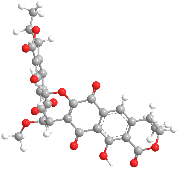
Secondary metabolites, 3D structure: Trivial name – xanthomegnin
Copyright: n/a
Notes:
Species: A. melleus, A. ochraceus, A. sulphureusSystematic name: (8,8′-Bi-1H-naphtho(1,2-c)pyran)-1,1′,7,7′,10,10′-hexone, 3,3′,4,4′-tetrahydro-6,6′-dihydroxy-9,9′-dimethoxy-3,3′-dimethyl-, (3R,3’R)- (8CI) (8,8′-BI-1H-NAPHTHO(2,3-c)PYRAN)-1,1′,6,6′,9,9′-HEXONE, 3,3′,4,4′-TETRAHYDRO-10, (8,8′-Bi-1H-naphtho(2,3-c)pyranMolecular formulae: C30H22O12Molecular weight: 574.488Chemical abstracts number: 1685-91-2Selected references: Durley RC, MacMillan J, Simpson TJ, Glen AT, Turner WB. Fungal products. Part XIII. Xanthomegnin, viomellin, rubrosulphin, and viopurpurin, pigments from Aspergillus sulphureus and Aspergillus melleus. J Chem Soc [Perkin 1]. 1975;(2):163-9. Stack ME, Mislivec PB. Production of xanthomegnin and viomellein by isolates of Aspergillus ochraceus, Penicillium cyclopium, and Penicillium viridicatum. Appl Environ Microbiol. 1978 Oct;36(4):552-4.Toxicity: Doses of 448 mg/kg body-weight in mice caused symptoms of liver damage including jaundice, necrotising cholangitis and other histological alterations. Robbers JE, Hong S, Tuite J, Carlton WW. Production of xanthomegnin and viomellein by species of Aspergillus correlated with mycotoxicosis produced in mice. Appl Environ Microbiol. 1978 Dec;36(6):819-23.
Images library
-
Title
Legend
-
Conidial head and brown conidia in a section of a fungus ball caused by Aspergillus niger (H&E, x 400).

-
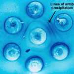
Double diffusion test for aspergillosis. Central well contains Aspergillus fumigatus antigen and wells in the top and bottom contain control antiserum.
-
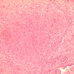
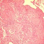
Allergic Bronchocentric Granulomatosis. low power. Sections show muscle, lung with acute inflammation and evidence of organisation with early fibrosis. The bronchial wall can be seen with chronic inflammation and many eosinophils.There is a thickened basement membrane. No definite granulomata are seen.

-
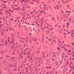
Allergic Bronchocentric Granulomatosis. Sections show muscle, lung with acute inflammation and evidence of organisation with early fibrosis. The bronchial wall can be seen with chronic inflammation and many eosinophils.There is a thickened basement membrane. No definite granulomata are seen.
-
Allergic Bronchocentric Granulomatosis. Higher power. Sections show muscle, lung with acute inflammation and evidence of organisation with early fibrosis. The bronchial wall can be seen with chronic inflammation and many eosinophils.There is a thickened basement membrane. No definite granulomata are seen.
-
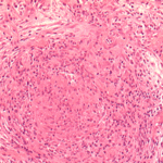
Allergic Bronchocentric Granulomatosis. Low power. Sections show muscle, lung with acute inflammation and evidence of organisation with early fibrosis. The bronchial wall can be seen with chronic inflammation and many eosinophils.There is a thickened basement membrane. No definite granulomata are seen.
-
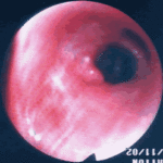
Allergic Bronchopulmonary Aspergillosis (ABPA). PT JC
CXR prior to bronchoscopy had shown an opacity just superior to the right hilum, which was felt to represent possibly a fungal plug. Patient was therefore bronchoscoped.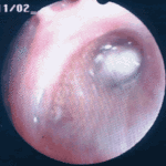 ,
, 
-
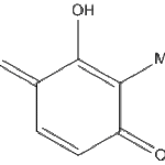
Secondary metabolites, Structural diagram. Trivial name – 2-hydroxy-3-methyl-1,4-benzoquinone
-
Secondary metabolite structure: trivial name – 13-O-Methylviriditin


