Date: 26 November 2013
Copyright: n/a
Notes:
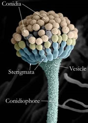
Aspergillus and other filamentous fungi produce clusters of spores known as fruiting bodies, which correspond with the mushrooms that other groups of fungi produce. They are very useful in diagnosing fungal diseases because their structures are strongly characteristic of different species.
Aspergillus was originally named for its fruiting body because it was thought that they resemble as aspergillum, which is a device used for sprinkling holy water.
In this picture, scanning electron microscopy has been used to visualise the fruiting body and false colour has been added to highlight the different parts.
Images library
-
Title
Legend
-
Conidial head and brown conidia in a section of a fungus ball caused by Aspergillus niger (H&E, x 400).

-
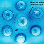
Double diffusion test for aspergillosis. Central well contains Aspergillus fumigatus antigen and wells in the top and bottom contain control antiserum.
-
Allergic Bronchocentric Granulomatosis. low power. Sections show muscle, lung with acute inflammation and evidence of organisation with early fibrosis. The bronchial wall can be seen with chronic inflammation and many eosinophils.There is a thickened basement membrane. No definite granulomata are seen.

-
Allergic Bronchocentric Granulomatosis. Sections show muscle, lung with acute inflammation and evidence of organisation with early fibrosis. The bronchial wall can be seen with chronic inflammation and many eosinophils.There is a thickened basement membrane. No definite granulomata are seen.
-
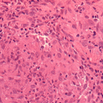
Allergic Bronchocentric Granulomatosis. Higher power. Sections show muscle, lung with acute inflammation and evidence of organisation with early fibrosis. The bronchial wall can be seen with chronic inflammation and many eosinophils.There is a thickened basement membrane. No definite granulomata are seen.
-
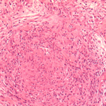
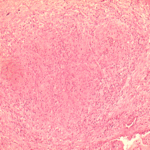
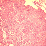
Allergic Bronchocentric Granulomatosis. Low power. Sections show muscle, lung with acute inflammation and evidence of organisation with early fibrosis. The bronchial wall can be seen with chronic inflammation and many eosinophils.There is a thickened basement membrane. No definite granulomata are seen.
-
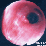
Allergic Bronchopulmonary Aspergillosis (ABPA). PT JC
CXR prior to bronchoscopy had shown an opacity just superior to the right hilum, which was felt to represent possibly a fungal plug. Patient was therefore bronchoscoped.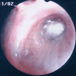 ,
, 
-
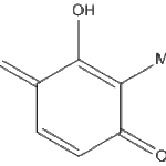
Secondary metabolites, Structural diagram. Trivial name – 2-hydroxy-3-methyl-1,4-benzoquinone
-
Secondary metabolite structure: trivial name – 13-O-Methylviriditin


