Date: 26 November 2013
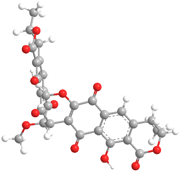
Secondary metabolites, 3D structure: Trivial name – xanthomegnin
Copyright: n/a
Notes:
Species: A. melleus, A. ochraceus, A. sulphureusSystematic name: (8,8′-Bi-1H-naphtho(1,2-c)pyran)-1,1′,7,7′,10,10′-hexone, 3,3′,4,4′-tetrahydro-6,6′-dihydroxy-9,9′-dimethoxy-3,3′-dimethyl-, (3R,3’R)- (8CI) (8,8′-BI-1H-NAPHTHO(2,3-c)PYRAN)-1,1′,6,6′,9,9′-HEXONE, 3,3′,4,4′-TETRAHYDRO-10, (8,8′-Bi-1H-naphtho(2,3-c)pyranMolecular formulae: C30H22O12Molecular weight: 574.488Chemical abstracts number: 1685-91-2Selected references: Durley RC, MacMillan J, Simpson TJ, Glen AT, Turner WB. Fungal products. Part XIII. Xanthomegnin, viomellin, rubrosulphin, and viopurpurin, pigments from Aspergillus sulphureus and Aspergillus melleus. J Chem Soc [Perkin 1]. 1975;(2):163-9. Stack ME, Mislivec PB. Production of xanthomegnin and viomellein by isolates of Aspergillus ochraceus, Penicillium cyclopium, and Penicillium viridicatum. Appl Environ Microbiol. 1978 Oct;36(4):552-4.Toxicity: Doses of 448 mg/kg body-weight in mice caused symptoms of liver damage including jaundice, necrotising cholangitis and other histological alterations. Robbers JE, Hong S, Tuite J, Carlton WW. Production of xanthomegnin and viomellein by species of Aspergillus correlated with mycotoxicosis produced in mice. Appl Environ Microbiol. 1978 Dec;36(6):819-23.
Images library
-
Title
Legend
-
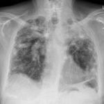
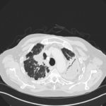
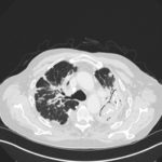
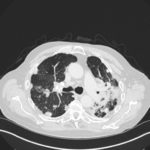
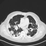
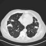
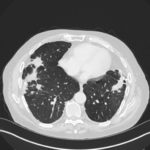
Mr RM is 80 and an ex-coal miner.He developed pneumoconiosis from exposure to coal dust. He also developed rheumatoid arthritis and the combination of this disease and pneumoconiosis is called Caplan’s syndrome.
His chest Xray in early 2015 shows extensive bilateral pulmonary shadowing with solid looking nodules superimposed on abnormal lung fields, contraction of his left lung with an elevated diaphragm and a large left upper lobe aspergilloma, displaying a classic air crescent. His CT scan from mid 2014 demonstrates a large aspergilloma in a cavity on the left, with marked pleural thickening around it, which is partially ‘calcified’ towards its base. Inferiorly on other images,remarkable pleural thickening and fibrotic irregular and spiculated nodules are seen, most partially calcified.
 ,
,  ,
,  ,
,  ,
,  ,
,  ,
, 