Date: 26 November 2013
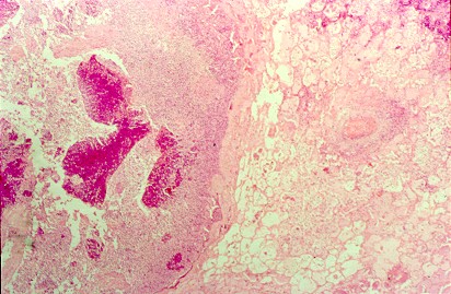
Pulmonary aspergillosis (cow 2). Section of lung from a 2 year old cow with weight loss and anorexia since calving. On necropsy examination, multiple firm masses were identified throughout the lungs. These were cavitating in nature, with a necrotic centre and peripheral fibrosis. Both this section and the following one are taken from the edge of such a lesion and demonstrate the pyogranulomatous inflammatory response.
Copyright:
© Dr. Michael Day, University of Bristol
Notes: n/a
Images library
-
Title
Legend
-
Conidial head and brown conidia in a section of a fungus ball caused by Aspergillus niger (H&E, x 400).

-
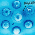
Double diffusion test for aspergillosis. Central well contains Aspergillus fumigatus antigen and wells in the top and bottom contain control antiserum.
-
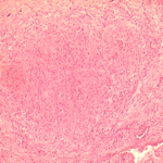
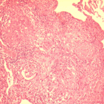
Allergic Bronchocentric Granulomatosis. low power. Sections show muscle, lung with acute inflammation and evidence of organisation with early fibrosis. The bronchial wall can be seen with chronic inflammation and many eosinophils.There is a thickened basement membrane. No definite granulomata are seen.

-
Allergic Bronchocentric Granulomatosis. Sections show muscle, lung with acute inflammation and evidence of organisation with early fibrosis. The bronchial wall can be seen with chronic inflammation and many eosinophils.There is a thickened basement membrane. No definite granulomata are seen.
-
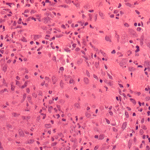
Allergic Bronchocentric Granulomatosis. Higher power. Sections show muscle, lung with acute inflammation and evidence of organisation with early fibrosis. The bronchial wall can be seen with chronic inflammation and many eosinophils.There is a thickened basement membrane. No definite granulomata are seen.
-
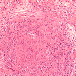
Allergic Bronchocentric Granulomatosis. Low power. Sections show muscle, lung with acute inflammation and evidence of organisation with early fibrosis. The bronchial wall can be seen with chronic inflammation and many eosinophils.There is a thickened basement membrane. No definite granulomata are seen.
-
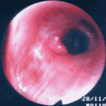
Allergic Bronchopulmonary Aspergillosis (ABPA). PT JC
CXR prior to bronchoscopy had shown an opacity just superior to the right hilum, which was felt to represent possibly a fungal plug. Patient was therefore bronchoscoped.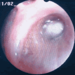 ,
, 
-
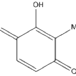
Secondary metabolites, Structural diagram. Trivial name – 2-hydroxy-3-methyl-1,4-benzoquinone
-
Secondary metabolite structure: trivial name – 13-O-Methylviriditin


