Date: 26 November 2013
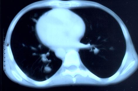
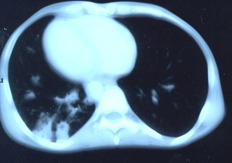
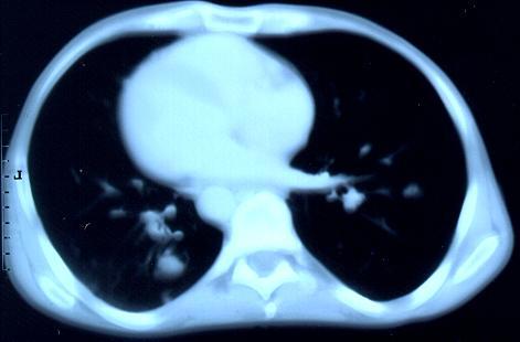
Transverse sections through the thorax of a patient with AIDS, hepatitis C and a left tempero-parietal cerebral lymphoma. His CD4 cell count was 45 x 106 / l. The lymphoma was proven by biopsy after a poor response to anti-toxoplasma therapy. He was given dexamethasone to cover the surgery and then developed diabetes mellitus. He did not receive chemotherapy for his lymphoma but did have 2 cerebral radiotherapy treatments (1.8 Gy each). Three weeks after the biopsy he developed dyspnoea and fever. Shortly after this he developed a right-sided hemiparesis, became comatose and died 2 days later.Autopsy showed a cerebral lymphoma and pulmonary and renal aspergillosis. Aspergillus nidulans was recovered from cultures of lungs and kidney.
Copyright:
Images submitted by Dr. Cornelia Lass-Floerl, University of Innsbruck – Institute of Hygiene; the case team includes: Dr. Mario Sarcletti, Dr. Alfons Stöger and Prof. Hans Maier all at the University of Innsbruck.
Notes: n/a
Images library
-
Title
Legend
-
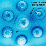
Double diffusion test for aspergillosis. Central well contains Aspergillus fumigatus antigen and wells in the top and bottom contain control antiserum.
-
Allergic Bronchocentric Granulomatosis. low power. Sections show muscle, lung with acute inflammation and evidence of organisation with early fibrosis. The bronchial wall can be seen with chronic inflammation and many eosinophils.There is a thickened basement membrane. No definite granulomata are seen.

-
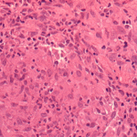
Allergic Bronchocentric Granulomatosis. Sections show muscle, lung with acute inflammation and evidence of organisation with early fibrosis. The bronchial wall can be seen with chronic inflammation and many eosinophils.There is a thickened basement membrane. No definite granulomata are seen.
-
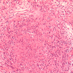
Allergic Bronchocentric Granulomatosis. Higher power. Sections show muscle, lung with acute inflammation and evidence of organisation with early fibrosis. The bronchial wall can be seen with chronic inflammation and many eosinophils.There is a thickened basement membrane. No definite granulomata are seen.
-
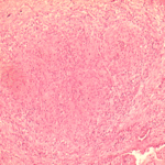
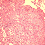
Allergic Bronchocentric Granulomatosis. Low power. Sections show muscle, lung with acute inflammation and evidence of organisation with early fibrosis. The bronchial wall can be seen with chronic inflammation and many eosinophils.There is a thickened basement membrane. No definite granulomata are seen.
-
Allergic Bronchopulmonary Aspergillosis (ABPA). PT JC
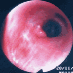
CXR prior to bronchoscopy had shown an opacity just superior to the right hilum, which was felt to represent possibly a fungal plug. Patient was therefore bronchoscoped.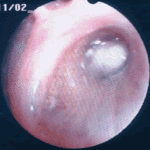 ,
, 
-
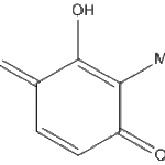
Secondary metabolites, Structural diagram. Trivial name – 2-hydroxy-3-methyl-1,4-benzoquinone
-
Secondary metabolite structure: trivial name – 13-O-Methylviriditin

-
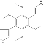
Secondary metabolites, structural diagram. Trivial name – 2”-oxoasterriquinol D Me ether 1
