Date: 26 November 2013
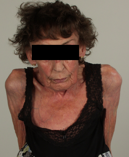
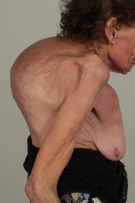
These pictures show remarkable curvature of the spine as a result of collapse of the vertebral bodies of the thoracic vertebrae. This is a gross example of steroid-induced osteoporosis. The dose was not large in the last 10 years, typically 5-10mg daily, but multiple high dose courses and slow tapering lead to this outcome.
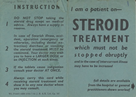
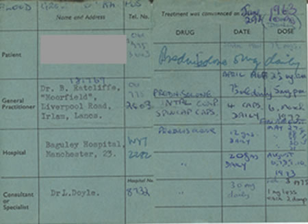
Her corticosteroid warning card is also demonstrated, as additional steroids are required for any significant illness or surgery, as her adrenal glands had completely atrophied.
Kindly supplied by Prof David Denning, South Manchester University Hospitals NHS Trust, Manchester UK
(© Fungal Research Trust)
Copyright:
Kindly supplied by Prof David Denning, South Manchester University Hospitals NHS Trust, Manchester UK
Notes:
Images library
-
Title
Legend
-
Conidial head and brown conidia in a section of a fungus ball caused by Aspergillus niger (H&E, x 400).

-
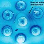
Double diffusion test for aspergillosis. Central well contains Aspergillus fumigatus antigen and wells in the top and bottom contain control antiserum.
-
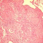
Allergic Bronchocentric Granulomatosis. low power. Sections show muscle, lung with acute inflammation and evidence of organisation with early fibrosis. The bronchial wall can be seen with chronic inflammation and many eosinophils.There is a thickened basement membrane. No definite granulomata are seen.

-
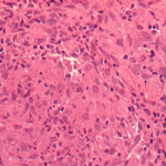
Allergic Bronchocentric Granulomatosis. Sections show muscle, lung with acute inflammation and evidence of organisation with early fibrosis. The bronchial wall can be seen with chronic inflammation and many eosinophils.There is a thickened basement membrane. No definite granulomata are seen.
-
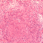
Allergic Bronchocentric Granulomatosis. Higher power. Sections show muscle, lung with acute inflammation and evidence of organisation with early fibrosis. The bronchial wall can be seen with chronic inflammation and many eosinophils.There is a thickened basement membrane. No definite granulomata are seen.
-
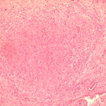
Allergic Bronchocentric Granulomatosis. Low power. Sections show muscle, lung with acute inflammation and evidence of organisation with early fibrosis. The bronchial wall can be seen with chronic inflammation and many eosinophils.There is a thickened basement membrane. No definite granulomata are seen.
-
Allergic Bronchopulmonary Aspergillosis (ABPA). PT JC
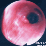
CXR prior to bronchoscopy had shown an opacity just superior to the right hilum, which was felt to represent possibly a fungal plug. Patient was therefore bronchoscoped.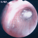 ,
, 
-
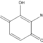
Secondary metabolites, Structural diagram. Trivial name – 2-hydroxy-3-methyl-1,4-benzoquinone
-
Secondary metabolite structure: trivial name – 13-O-Methylviriditin


