Date: 26 November 2013
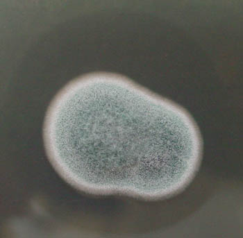
Colony on malt agar (approx 12 mm diameter)
Copyright:
Images were kindly provided by Niall Hamilton, 09/05. Copyright Fungal Research Trust
Notes:
Cultures were grown on malt extract agar at 30°C for 6 days
Images library
-
Title
Legend
-
Conidial head and brown conidia in a section of a fungus ball caused by Aspergillus niger (H&E, x 400).

-
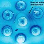
Double diffusion test for aspergillosis. Central well contains Aspergillus fumigatus antigen and wells in the top and bottom contain control antiserum.
-
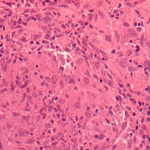
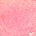
Allergic Bronchocentric Granulomatosis. low power. Sections show muscle, lung with acute inflammation and evidence of organisation with early fibrosis. The bronchial wall can be seen with chronic inflammation and many eosinophils.There is a thickened basement membrane. No definite granulomata are seen.

-
Allergic Bronchocentric Granulomatosis. Sections show muscle, lung with acute inflammation and evidence of organisation with early fibrosis. The bronchial wall can be seen with chronic inflammation and many eosinophils.There is a thickened basement membrane. No definite granulomata are seen.
-
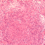
Allergic Bronchocentric Granulomatosis. Higher power. Sections show muscle, lung with acute inflammation and evidence of organisation with early fibrosis. The bronchial wall can be seen with chronic inflammation and many eosinophils.There is a thickened basement membrane. No definite granulomata are seen.
-
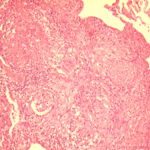
Allergic Bronchocentric Granulomatosis. Low power. Sections show muscle, lung with acute inflammation and evidence of organisation with early fibrosis. The bronchial wall can be seen with chronic inflammation and many eosinophils.There is a thickened basement membrane. No definite granulomata are seen.
-
Allergic Bronchopulmonary Aspergillosis (ABPA). PT JC
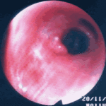
CXR prior to bronchoscopy had shown an opacity just superior to the right hilum, which was felt to represent possibly a fungal plug. Patient was therefore bronchoscoped.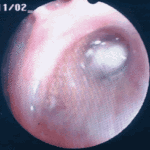 ,
, 
-
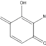
Secondary metabolites, Structural diagram. Trivial name – 2-hydroxy-3-methyl-1,4-benzoquinone
-
Secondary metabolite structure: trivial name – 13-O-Methylviriditin


