Date: 3 May 2013
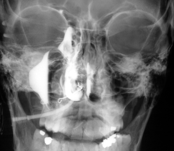
Yamik catheter for rinsing nasal and paranasal cavities. Image D
Copyright:
These images were kindly given by Andreas Neher of the Department of Otorhinolaryngology, Innsbruck Medical University, Anichstr. 35, Innsbruck, Austria. For further details of the procedure please refer to the PDF link above. (C) Fungal Research Trust
Notes:
Rinsing of the nasal and paranasal cavities applied via a novel catheter system (YAMIK). The cavities were rinsed with 10–20 ml of 1% aqueous N-Chlorotaurine (NCT) solution, treatment consisted of three lavages per week for 4 weeks. N-Chlorotaurine is a mild endogenous oxidant with broad-spectrum antimicrobial activity. The use of the catheter proved to be a successful way of rinsing the nasal and paranasal cavities. Link to PDF with further details.
C,D X-ray set up of a healthy volunteer rinsed with 10 ml of radio-opaque solution via YAMIK catheter demonstrating sufficient filling of the paranasal sinuses.
Images library
-
Title
Legend
-
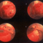
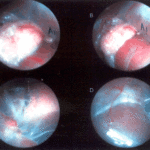
Falcons: The following images were obtained by endoscopy of falcons with aspergillosis.A,B Thoracic airsac (T) with prominent blood vessels and a dead serratospiculum worm (W). The presence of these lung worms makes the airsac look milky. D Normal ovary with developing follicles.

-
Falcons: The following images were obtained by endoscopy of falcons with aspergillosis.B,D Aspergillus lesions (A) over a swollen liver

-
Falcons: The following images were obtained by endoscopy of falcons with aspergillosis.B Cranial, middle, caudal lobes (K1,K2,K3) of the left kidney, all the lobes show slight nephromegaly.C Yellow aspergillus colony (A1), lying adjacent to the lung.D White aspergillus colonies (A2,A3,A4).
-
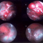
Falcons: The following images were obtained by endoscopy of falcons with aspergillosis.C Cranial pole of left kidney (K) -mildly inflamed.D Ovary ( F) with developing follicles.
-
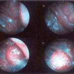
The following images were obtained by endoscopy of falcons with aspergillosis.A and B Lung Worm (S) over liver (Li) (serratospiculum seurati)C and D Aspergilloma (A) and prominent blood vessels on the caudal thoracic air sac (T).
