Date: 7 February 2014
Copyright: n/a
Notes:
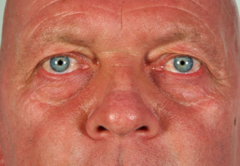
This man with immunocompromised with autoimmune disease developed bilateral invasive aspergillosis and a galactomannan antigen OD in BAL of 9.0. He was started on voriconazole and responded well. Some weeks later his face became erythematous and slightly uncomfortable. The photographs show the remarkable extent of his voriconazole photosensitivity with very little conjunctivitis or cheilitis (lip dryness). Monochromator testing to narrow band UVB, UVA and visible light and provocation testing was within normal limits. Voriconazole was stopped after 9 months of therapy and reduction of immunosuppression, with resolution of photosensitivity.
Images library
-
Title
Legend
-
1 Axial computed tomography (CT) scans of the frontal sinus.
A: due to the long lasting pressure of mucus, the bone of the anterior wall of frontal sinus is thinned out and elevated anteriorly, forming a bulge. B: same situation as depicted in fig A: the posterior bony wall of frontal sinus is thinned out and extremely elevated posteriorly towards the frontal lobe of the brain. As depicted on the scan, a thin bony layer covering the dura could be recognized intraoperatively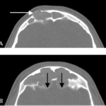
-
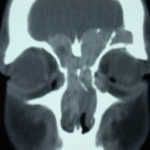
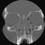
2 Same patient as 1 and 3, frontal CT
-
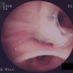
D. 6 months later, tenacious yellow secretions in L basal bronchial division
-
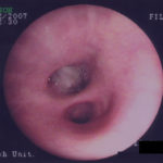
C. After suction the material was seen to extend distally – obstructing the right basal stem bronchus
-
B. After suction the material was seen to extend distally – obstructing the right basal stem bronchus
-
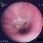
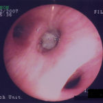
A. Necrotic mass prolapsing in and out of the distal right intermediate bronchus obscuring both the basal stem and basal division
