Date: 26 November 2013
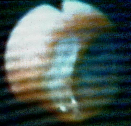
Bronchoscopic view of a deep bronchial ulcer in a lung transplant patient. Biopsies through the ulcer yielded cartilage with hyphae invading it. Fungal cultures of bronchial lavage grew Aspergillus fumigatus. He responded to oral itraconazole therapy.
Copyright: n/a
Notes:
This patient was reported in Kramer MR, Denning DW, Marshall SE, Ross D, Berry G, Lewiston N, Stevens DA, Theodore J. Ulcerative tracheobronchitis following lung transplantation: a new form of invasive aspergillosis. Am Rev Resp Dis 1991; 144: 552-556.
Images library
-
Title
Legend
-
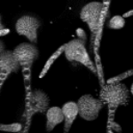
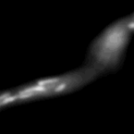
Mitochondria organisation: GFP fluorescence micrographs showing mitochondrial organisation in an A.nidulans strain with GFP mitochondria, grown at 25°C in minimal media.
-
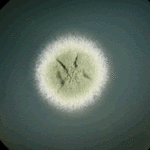
Colony morphology of A.nidulans SRF200 after two days at 37°C
-
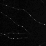
Aspergillus nidulans. Cell nuclei-Ds red. DsRed fluorescence micrographs showing nuclear distribution in an A.nidulans germling with dsRed stained nuclei

-
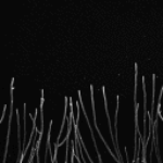
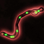
Cell Biology – Aspergillus nidulans. Cell nuclei-GFP. Nuclear distribution: GFP fluorescence mirographs showing fungal cell morphology and nuclear distribution in A.nidulans. GFP stained nuclei,grown at 25°C in minimal media O/N
-
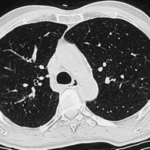
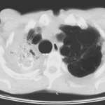
High resolution CT scan of chest.CT scan demonstrating remarkable bronchial wall thickening of the right main bronchus and main branches, in context of longstanding ABPA


