Date: 26 November 2013
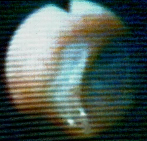
Bronchoscopic view of a deep bronchial ulcer in a lung transplant patient. Biopsies through the ulcer yielded cartilage with hyphae invading it. Fungal cultures of bronchial lavage grew Aspergillus fumigatus. He responded to oral itraconazole therapy.
Copyright: n/a
Notes:
This patient was reported in Kramer MR, Denning DW, Marshall SE, Ross D, Berry G, Lewiston N, Stevens DA, Theodore J. Ulcerative tracheobronchitis following lung transplantation: a new form of invasive aspergillosis. Am Rev Resp Dis 1991; 144: 552-556.
Images library
-
Title
Legend
-
Image A
CT Scan 30/3/99
Showing extreme pleural thickening and 2 small cavities at apex of left lung.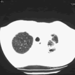
-
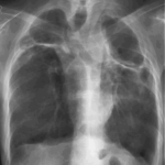
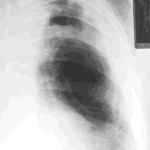
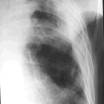
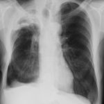
A 43 year old with smoking related emphysema was admitted to hospital with two separate episodes of haemoptysis. He had been in good health up to 1989, when he was diagnosed as having bilateral pulmonary tuberculosis. At that time a CT scan revealed a cavity in the left upper lobe (20.8cm2) with adjacent confluent infiltrates and pleural thickening. On bronchoscopic examination no abnormalities were noted and endobronchial biopsies did not reveal hyphae.
Over the next 4 years his condition deteriorated and a CT scan showed the left upper lobe cavity had increased to 40cm2. Itraconazole 400mg daily was prescribed. There was some clinical improvement on itraconazole but patient eventually deteriorated with breathlessness and with significant weight loss.
 ,
, 