Date: 26 November 2013
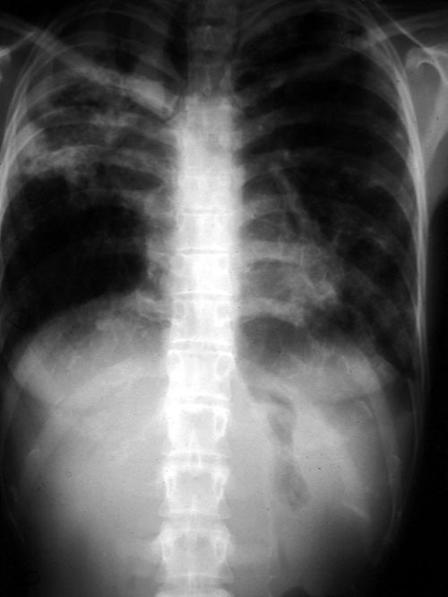
Image A. This 25 year old woman was previously well and presented with a pneumonia of uncertain aetiology. She has infiltrates in right upper-lobe and left middle and lower zones. The diagnosis was later made of chronic invasive pulmonary aspergillosis by bronchoscopy . Subsequently she was diagnosed with adult-onset chronic granulomatous disease with neutrophil function assays.
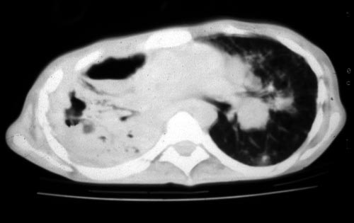
Image B. CT scan of the thorax just below the carina, showing almost complete opacification of the right lung and marked nodular shadowing around the hilum of the left lung.
Image C. Progression of pulmonary infiltrates are seen seven weeks later, despite administration of amphotericin B.
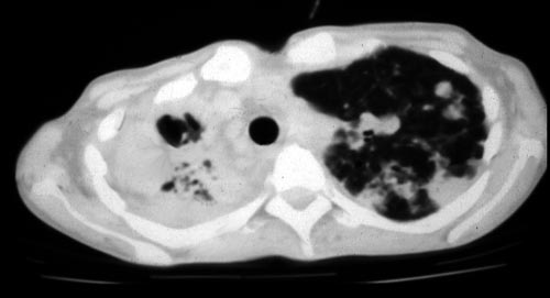
Image D. CT scan of the thorax above the carina showing near complete opacification of the right lung and multiple discrete nodular shadows in the left lung.
Copyright: n/a
Notes: n/a
Images library
-
Title
Legend
-
Culture plates on different media. A Colonies on CZ at 24oC, B on CYA at 20oC C on SAB at 37oC, D on CYA at 24oC

-
Mucous containing Charcot-Leyden crystals, stained with H & EA 57 year old woman presented with breathlessness. She had a history of mild asthma for which she occasionally took salbutamol inhaler puffs. The patient underwent a pneumonectomy because of the severity of her disease process, and uncertainty about the diagnosis, prior to serology results being obtained.Serology showed an IgE of 2600, with a strongly positive Aspergillus RAST test and weakly positive Aspergillus precipitins. Material re


