Date: 26 November 2013
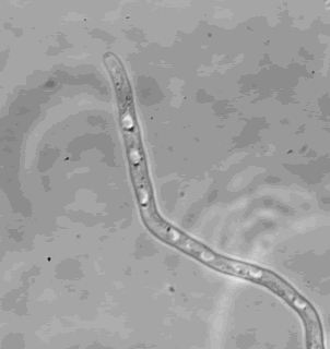
Spitzenkorper; phase contrast micrograph of A.nidulans after one day grown at 25°C in minimal media containing 17% gelatin showing spitzenkorper and fungal cell morphology
Copyright:
Kindly provided by Sven Konzack and Reinhard Fischer Max-Planck-Institute for Terrestrial Microbiology, Karl von Frisch Str, D 35043 Marburg, Germany.
Notes: n/a
Images library
-
Title
Legend
-
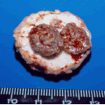
Gross pathology demonstrating the great pleural thickness and two cavities (upper lobe and superior segment of lower lobe) with fragments of fungal mass.

-
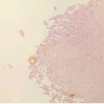
Histopathological appearance of a fungus ball. Note a conidial head resulting from fungal exposure to the air.
-
Histopathological appearance of a fungus ball caused by Scedosporium apiospermum. The presence of anneloconidia differentiates it from Aspergillus.

-
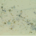
Chronic necrotising aspergillosis. Hyaline hyphal and calcium oxalate crystals obtained by needle aspirate biopsy from a diabetic patient with chronic necrotizing aspergillosis caused by Aspergillus niger (Papanicolaou, x 100).
-
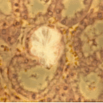
Aspergillus niger fungus ball and acute oxalosis. Higher magnification of adjacent replicate section.
-
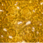
Oxalate crystals within renal tubuli (H&E, phase contrast, x 100). This patient developed acute oxalosis.
-
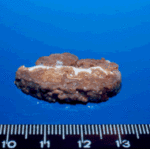
Lung surface. Fungus ball, severe parenchymal fibrosis and pleural thickening.

-
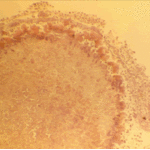
The periphery of the fungus ball is deeply eosinophilic because of the deposition of Splendore-Hoeppli material.

 ,
, 
 ,
,  ,
, 