Date: 3 April 2014
Copyright: n/a
Notes:
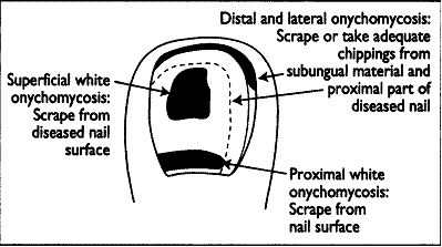
The quality of the specimen taken is a major factor in success or otherwise of microscopy and culture. Having a specimen taken should be painless apart from occasional slight discomfort when subungual specimens are taken. The figure shows the appropriate sites from which nail specimens should be obtained.
Images library
-
Title
Legend
-
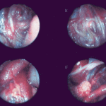
Falcons: The following images were obtained by endoscopy of falcons with aspergillosis.A,B Thoracic airsac (T) with prominent blood vessels and a dead serratospiculum worm (W). The presence of these lung worms makes the airsac look milky. D Normal ovary with developing follicles.

-
Falcons: The following images were obtained by endoscopy of falcons with aspergillosis.B,D Aspergillus lesions (A) over a swollen liver

-
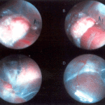
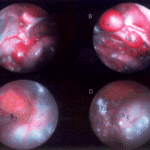
Falcons: The following images were obtained by endoscopy of falcons with aspergillosis.B Cranial, middle, caudal lobes (K1,K2,K3) of the left kidney, all the lobes show slight nephromegaly.C Yellow aspergillus colony (A1), lying adjacent to the lung.D White aspergillus colonies (A2,A3,A4).
-
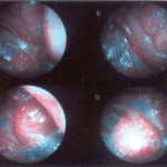
Falcons: The following images were obtained by endoscopy of falcons with aspergillosis.C Cranial pole of left kidney (K) -mildly inflamed.D Ovary ( F) with developing follicles.
-
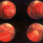
The following images were obtained by endoscopy of falcons with aspergillosis.A and B Lung Worm (S) over liver (Li) (serratospiculum seurati)C and D Aspergilloma (A) and prominent blood vessels on the caudal thoracic air sac (T).
-
The following images were obtained by endoscopy of falcons with aspergillosis. A Lung Worm (serratospiculum sp.) B Lying in betweeen loops of the intestine.C An aspergillus lesion in between the loops of the intestineD Showing cranial pole of left kidney, ovary and oviduct.
