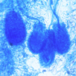
Date: 26 November 2013
D Sinusitis radiology with fluid level
Copyright:
Fungal Infection Trust
Notes:
Bronchography (A & B)is an old technique for visualising the bronchial tree, by introducing radio-opaque dye into the airways and then taking a chest Xray. It was the first means used to diagnose bronchiectasis, seen in these examples below. An historical description of the technique from 1922 can be seen here
Nowadays CT scanning of the chest is used for this purpose with 3D reconstruction in some cases.
White cell scan (C) This pair of white cell scans from different people show almost no white cells in the lungs on the left, as in a healthy person (the spleen is the ‘hottest area). The scan on the right shows grossly increased update, especially in the left lung (seen on the right). This is the typical feature of severe bronchiectasis with large amounts of neutrophils and other phagocytes present.
Sinusitis Plain X-ray (D) of the face showing the maxillary sinuses. The right maxillary sinus is complete fluid filled and the left side (seen on the right) has a fluid level. These features may be seen with severe acute bacterial sinusitis, but also other causes of sinusitis, including allergic rhinosinusitis.
Images library
-
Title
Legend
-
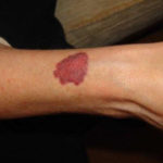
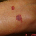
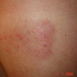
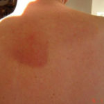
Drug rashes: Drug interactions between steroids and anti-fungal drugs – (ecchymosis)
 ,
,  ,
,  ,
, 
-
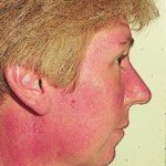
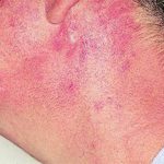
Reference: Muco-cutaneous retinoid effects and facial erythema related to the novel triazole antifungal agent voriconazole. Denning, DW & Griffiths, CEM. Clin.Exp Dermatol 2001, 26(8), 648-53.
Courtesy of Dr D Denning, Wythenshawe Hospital, Manchester.(© Fungal Research Trust)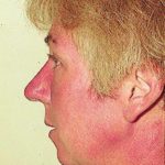 ,
,  ,
, 
-
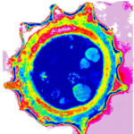
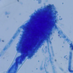
Micrographs of A. niger conidia & conidial heads provided by Amaliya Stepanova, Head of Laboratory pathomorphology and cytology at Kashkin Research Institute, Russian Federation.
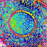 ,
, 
-
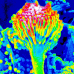
Micrographs of A. terreus conidia & conidial heads provided by Amaliya Stepanova, , Head of Laboratory pathomorphology and cytology at Kashkin Research Institute, Russian Federation.
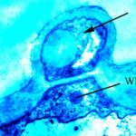 ,
,  ,
, 
-
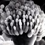
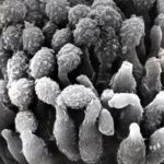
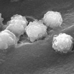
Micrographs of A. fumigatus conidia & conidial heads provided by Amaliya Stepanova, , Head of Laboratory pathomorphology and cytology at Kashkin Research Institute, Russian Federation.
 ,
,  ,
,  ,
,  ,
, 
-
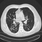
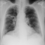
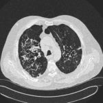
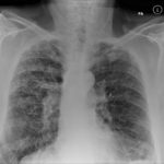
Patients has history of ABPA complicating long standing asthma. His total IgE has fluctuated between 2,200 and 4,600 KU/L, his Aspergillus IgE between 36.3 and 65.4 kAU/L and Aspergillus IgG from 87-154 mg/L. He has been taking long term itraconazole.
 ,
,  ,
,  ,
, 

 ,
,  ,
, 