Date: 26 November 2013
D Sinusitis radiology with fluid level
Copyright:
Fungal Infection Trust
Notes:
Bronchography (A & B)is an old technique for visualising the bronchial tree, by introducing radio-opaque dye into the airways and then taking a chest Xray. It was the first means used to diagnose bronchiectasis, seen in these examples below. An historical description of the technique from 1922 can be seen here
Nowadays CT scanning of the chest is used for this purpose with 3D reconstruction in some cases.
White cell scan (C) This pair of white cell scans from different people show almost no white cells in the lungs on the left, as in a healthy person (the spleen is the ‘hottest area). The scan on the right shows grossly increased update, especially in the left lung (seen on the right). This is the typical feature of severe bronchiectasis with large amounts of neutrophils and other phagocytes present.
Sinusitis Plain X-ray (D) of the face showing the maxillary sinuses. The right maxillary sinus is complete fluid filled and the left side (seen on the right) has a fluid level. These features may be seen with severe acute bacterial sinusitis, but also other causes of sinusitis, including allergic rhinosinusitis.
Images library
-
Title
Legend
-
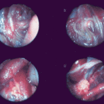
Falcons: The following images were obtained by endoscopy of falcons with aspergillosis.A,B Thoracic airsac (T) with prominent blood vessels and a dead serratospiculum worm (W). The presence of these lung worms makes the airsac look milky. D Normal ovary with developing follicles.

-
Falcons: The following images were obtained by endoscopy of falcons with aspergillosis.B,D Aspergillus lesions (A) over a swollen liver

-
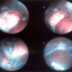
Falcons: The following images were obtained by endoscopy of falcons with aspergillosis.B Cranial, middle, caudal lobes (K1,K2,K3) of the left kidney, all the lobes show slight nephromegaly.C Yellow aspergillus colony (A1), lying adjacent to the lung.D White aspergillus colonies (A2,A3,A4).
-
Falcons: The following images were obtained by endoscopy of falcons with aspergillosis.C Cranial pole of left kidney (K) -mildly inflamed.D Ovary ( F) with developing follicles.
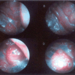
-
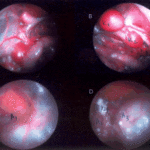
The following images were obtained by endoscopy of falcons with aspergillosis.A and B Lung Worm (S) over liver (Li) (serratospiculum seurati)C and D Aspergilloma (A) and prominent blood vessels on the caudal thoracic air sac (T).
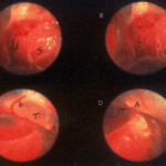
-
The following images were obtained by endoscopy of falcons with aspergillosis. A Lung Worm (serratospiculum sp.) B Lying in betweeen loops of the intestine.C An aspergillus lesion in between the loops of the intestineD Showing cranial pole of left kidney, ovary and oviduct.
