Date: 26 November 2013
D Sinusitis radiology with fluid level
Copyright:
Fungal Infection Trust
Notes:
Bronchography (A & B)is an old technique for visualising the bronchial tree, by introducing radio-opaque dye into the airways and then taking a chest Xray. It was the first means used to diagnose bronchiectasis, seen in these examples below. An historical description of the technique from 1922 can be seen here
Nowadays CT scanning of the chest is used for this purpose with 3D reconstruction in some cases.
White cell scan (C) This pair of white cell scans from different people show almost no white cells in the lungs on the left, as in a healthy person (the spleen is the ‘hottest area). The scan on the right shows grossly increased update, especially in the left lung (seen on the right). This is the typical feature of severe bronchiectasis with large amounts of neutrophils and other phagocytes present.
Sinusitis Plain X-ray (D) of the face showing the maxillary sinuses. The right maxillary sinus is complete fluid filled and the left side (seen on the right) has a fluid level. These features may be seen with severe acute bacterial sinusitis, but also other causes of sinusitis, including allergic rhinosinusitis.
Images library
-
Title
Legend
-
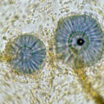
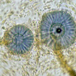
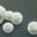
Scanning electron micrograph of Aspergillus ochraceopetaliformis conidial heads
-
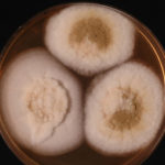
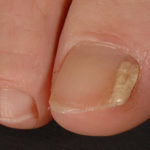
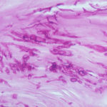
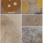
Image D & E. A case of onychomycosis associated with Aspergillus ochraceopetaliformis as described in Nail infection by Aspergillus ochraceopetaliformis. Med Mycol. 2009 Mar 9:1-5, 2009, Brasch J, Varga J, Jensen JM, Egberts F & Tintelnot K
 ,
,  ,
,  ,
,  ,
, 
-
Further details
Image 5. Oral itraconazole pulse therapy was given to the patient (200 mg twice daily for 1 week, with 3 weeks off between successive pulses, for four pulses) and treatment was successful.
 ,
,  ,
,  ,
,  ,
, 
-
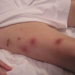
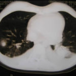
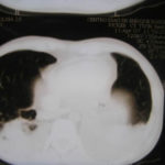
This patient was 28 yr old with adult lymphocytic leukaemia. She received induction chemotherapy and this infection developed 2 days after recovering from neutropenia.
 ,
,  ,
,  ,
,  ,
,  ,
,  ,
,  ,
,  ,
,  ,
, 
-
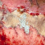
Close-up image of the lesion on the left thigh showing a mat of hyphae over the wound.
-
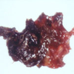
Eosinophilic mucin with A. flavus in the nasal cavity. Irregular crust of 2.5 cm from a patient diagnosed as allergic fungal sinusitis. Patient with allergic fungal sinusitis
-
GMS stain of eosinophilic mucin reveals a darkly stained dichotomously branched A. flavus hyphae within cellular background. Patient with allergic fungal sinusitis
