Date: 26 November 2013
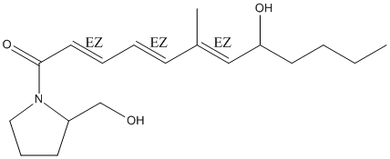
Secondary metabolites, structure diagram: Trivial name – Viriditin
Copyright: n/a
Notes:
Species: A. viridinutansSystematic name: 2-Pyrrolidinemethanol, 1-[(2E,4E,6E)-8-hydroxy-6-methyl-1-oxo-2,4,6-dodecatrienyl]-, (-)-Molecular formulae: C18H29NO3Molecular weight: 307Chemical abstracts number: 287102-33-4Selected references: Omolo, Josiah Ouma; Anke, Heidrun; Chhabra, Sumesh; Sterner, Olov (CORPORATE SOURCE Department of Biotechnology, University of Kaiserslautern, Kaiserslautern D-67663, Germany). SOURCE J. Nat. Prod., 63(7), 975-977 (English) 2000 American Chemical Society
Images library
-
Title
Legend
-
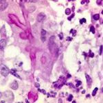
BAL specimen showing hyaline, septate hyphae consistent with Aspergillus, stained with Blankophor
-
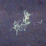
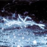
Mucous plug examined by light microscopy with KOH, showing a network of hyaline branching hyphae typical of Aspergillus, from a patient with ABPA.
-
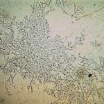
Corneal scraping stained with lactophenol cotton blue showing beaded septate hyphae not typical of either Fusarium spp or Aspergillus spp, being more consistent with a dematiceous (ie brown coloured) fungus
-
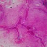
Corneal scrape with lactophenol cotton blue shows separate hyphae with Fusarium spp or Aspergillus spp.
-
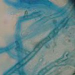
A filamentous fungus in the CSF of a patient with meningitis that grew Candida albicans in culture subsequently.
-
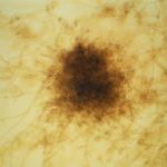
Transmission electron micrograph of a C. neoformans cell seen in CSF in an AIDS patients with remarkably little capsule present. These cells may be mistaken for lymphocytes.
-
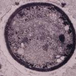
India ink preparation of CSF showing multiple yeasts with large capsules, and narrow buds to smaller daughter cells, typical of C. neoformans
