Date: 26 November 2013
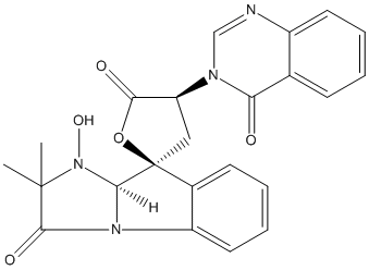
Secondary metabolites, structure diagram: Trivial name – tryptoquivaline L
Copyright: n/a
Notes:
Species: A. fumigatusSystematic name: Spiro[furan-2(5H),9′-[9H]imidazo[1,2-a]indole]-3′,5(2’H)-dione, 1′,3,4,9’a-tetrahydro-1′-hydroxy-2′,2′-dimethyl-4-(4-oxo-3(4H)-quinazolinyl)-, (2S,4S,9’aS)-Molecular formulae: C23H20N4O5Molecular weight: 432Chemical abstracts number: 69483-20-1Selected references: Yamazaki, Mikio; Fujimoto, Haruhiro; Maebayashi, Yukio; Okuyama, Emi (Res. Inst. Chemobiodyn., Chiba Univ., Chiba, Japan). Tennen Yuki Kagobutsu Toronkai Koen Yoshishu, 21st, 14-21. Hokkaido Daigaku Nogakubu: Sapporo, Japan. (Japanese) 1978.
Images library
-
Title
Legend
-
Further details
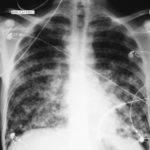
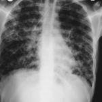
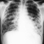
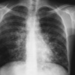
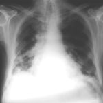
Image 1. The chest x-ray shows extensive bilateral nodular disease, most consistent with a fungal infection, or possibly tuberculosis. He was treated with a bucket face mask with 80% oxygen and voriconazole.
 ,
,  ,
,  ,
,  ,
,  ,
, 
-
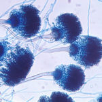
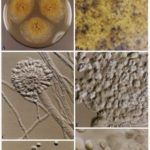
A Colonies on MEA +20 % sucrose after 2 weeks; B ascomata, x 40; C conidiophore of Aspergillus glaucus x 920;D conidiophore of Aspergillus glaucus x920 E. portion of ascoma with asci x 920. F ascospores x2330.
-
Scanning electron micrographs of A. fumigatus conidia of transformants rodB-02 (b). Size bar, 100 nm.

-
Scanning electron micrographs of A. fumigatus conidia of the wild-type G10 strain (a). Size bar, 100 nm.
-
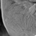
Scanning electron micrographs of A. fumigatus conidia of rodA rodB-26 (d).Size bar, 100 nm.
-
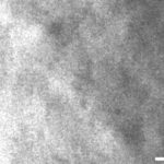
Scanning electron micrograph of an A.fumigatus conidium of rodA-47 (c), showing the hydrophobic rodlets covering the surface. Size bar, 100 nm.

 ,
, 
