Date: 26 November 2013
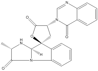
Secondary metabolites, structure diagram: Trivial name – tryptoquivaline J
Copyright: n/a
Notes:
Species: A. fumigatusSystematic name: Spiro[furan-2(5H),9′-[9H]imidazo[1,2-a]indole]-3′,5(2’H)-dione, 1′,3,4,9’a-tetrahydro-2′-methyl-4-(4-oxo-3(4H)-quinazolinyl)-, [2’S-[2’a,9’b(S*),9’aa]]-Molecular formulae: C22H18N4O4Molecular weight: 402.403Chemical abstracts number: 66212-51-9Selected references: Yamazaki, Mikio; Fujimoto, Haruhiro; Okuyama, Emi (Res. Inst. Chemobiodyn., Chiba Univ., Chiba, Japan). Chem. Pharm. Bull., 26(1), 111-17 (English) 1978.SECONDARY METABOLITES MYCOTOXINS PRODUCED BY FUNGI COLONIZING CEREAL GRAIN IN STORAGE STRUCTURE AND PROPERTIES GOLINSKI P CHELKOWSKI, J. (ED.). DEVELOPMENTS IN FOOD SCIENCE, VOL. 26. CEREAL GRAIN: MYCOTOXINS, FUNGI AND QUALITY IN DRYING AND STORAGE. XXII+607P. ELSEVIER SCIENCE PUBLISHERS B.V.: AMSTERDAM, NETHERLANDS; (DIST. IN THE USA AND CANADA BY ELSEVIER SCIENCE PUBLISHING CO., INC.: NEW YORK, NEW YORK, USA). ILLUS. MAPS. ISBN 0-444-88554-4.; 0 (0). 1991. 355-403.
Images library
-
Title
Legend
-
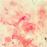
Corneal ulcer – gram stain. Corneal scrapings were taken from a 67 yr old farmer presenting with a corneal ulcer of the right eye. A piece of vegetable matter was embedded in the cornea and scrapings were done. Gram stain (500x magnification) showed numerous septate hyphae. Cultures grew a small amount of A fumigatus.

-
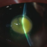
Aspergillus keratitis. Central lesion in aspergillus keratitis following a corneal foreign body which made a good response to topical treatment alone, albeit over 2 months intensive treatment.
-
Aspergillus keratitis. B- Severe central aspergillus infection with a “cheesey†looking area of the lesion and hypopyon (fluid level of inflammatory cells in the anterior chamber)
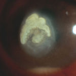
-
Aspergillus keratitis. A- Severe aspergillus infection with large area of corneal ulceration and deep stromal involvement
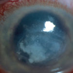
-
Candida keratitis. Focal candida keratitis as an unusual cause of a suture related infection following corneal transplantation for non infective indication
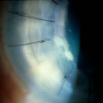
-
Candida keratitis. Subacute onset of candida keratitis in a young adult in whom dust blew into her eye in Greece. A slightly “feathery†edge to stromal involvement is suggestive of fungal infection
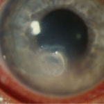
-
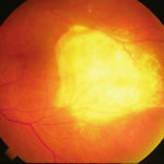
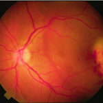
Aspergillus endopthalmitis. Temporal necrosis due to Aspergillus endopthalmitis as part of disseminated disease. No evidence of vitritis. Systemic treatment essential.
-
Aspergillus endopthalmitis. Large scarred area of the choroid following healing after Aspergillus endopthalmitis
