Date: 26 November 2013
Secondary metabolites, structure diagram: Trivial name – tryptoquivaline H
Copyright: n/a
Notes:
Species: A. fumigatusSystematic name: Spiro[furan-2(5H),9′-[9H]imidazo[1,2-a]indole]-3′,5(2’H)-dione, 1′,3,4,9’a-tetrahydro-1′-hydroxy-2′-methyl-4-(4-oxo-3(4H)-quinazolinyl)-, (2S,2’S,4S,9’aS)-Molecular formulae: C22H18N4O5Molecular weight: 418.402Chemical abstracts number: 61949-67-5Selected references: Yamazaki, Mikio; Fujimoto, Haruhiro; Okuyama, Emi (Res. Inst. Chemobiodynamics, Chiba Univ., Chiba, Japan). Tetrahedron Lett., (33), 2861-4 (English) 1976.SECONDARY METABOLITES MYCOTOXINS PRODUCED BY FUNGI COLONIZING CEREAL GRAIN IN STORAGE STRUCTURE AND PROPERTIES GOLINSKI P CHELKOWSKI, J. (ED.). DEVELOPMENTS IN FOOD SCIENCE, VOL. 26. CEREAL GRAIN: MYCOTOXINS, FUNGI AND QUALITY IN DRYING AND STORAGE. XXII+607P. ELSEVIER SCIENCE PUBLISHERS B.V.: AMSTERDAM, NETHERLANDS; (DIST. IN THE USA AND CANADA BY ELSEVIER SCIENCE PUBLISHING CO., INC.: NEW YORK, NEW YORK, USA). ILLUS. MAPS. ISBN 0-444-88554-4.; 0 (0). 1991. 355-403.
Images library
-
Title
Legend
-
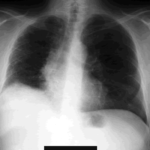
The chest x-ray shows a patient who had a left lung transplanted in May 2003 for cryptogenic fibrosing alveolitis, which was diagnosed post-transplant as sarcoidosis.
-
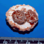
Gross pathology demonstrating the great pleural thickness and two cavities (upper lobe and superior segment of lower lobe) with fragments of fungal mass.

-
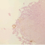
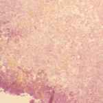
Histopathological appearance of a fungus ball. Note a conidial head resulting from fungal exposure to the air.
-
Histopathological appearance of a fungus ball caused by Scedosporium apiospermum. The presence of anneloconidia differentiates it from Aspergillus.

-
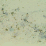
Chronic necrotising aspergillosis. Hyaline hyphal and calcium oxalate crystals obtained by needle aspirate biopsy from a diabetic patient with chronic necrotizing aspergillosis caused by Aspergillus niger (Papanicolaou, x 100).
-
Aspergillus niger fungus ball and acute oxalosis. Higher magnification of adjacent replicate section.
-
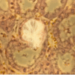
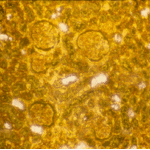
Oxalate crystals within renal tubuli (H&E, phase contrast, x 100). This patient developed acute oxalosis.
-
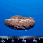
Lung surface. Fungus ball, severe parenchymal fibrosis and pleural thickening.


 ,
, 
 ,
,  ,
, 