Date: 26 November 2013
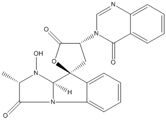
Secondary metabolites, structure diagram: Trivial name – tryptoquivaline E
Copyright: n/a
Notes:
Species: A. fumigatusSystematic name: Spiro[furan-2(5H),9′-[9H]imidazo[1,2-a]indole]-3′,5(2’H)-dione, 1′,3,4,9’a-tetrahydro-1′-hydroxy-2′-methyl-4-(4-oxo-3(4H)-quinazolinyl)-, (2S,2’S,4R,9’aS)-Molecular formulae: C22H18N4O5Molecular weight: 418.402Chemical abstracts number: 61897-87-8Selected references: Yamazaki, Mikio; Fujimoto, Haruhiro; Okuyama, Emi (Res. Inst. Chemobiodynamics, Chiba Univ., Chiba, Japan). Tetrahedron Lett., (33), 2861-4 (English) 1976.SECONDARY METABOLITES MYCOTOXINS PRODUCED BY FUNGI COLONIZING CEREAL GRAIN IN STORAGE STRUCTURE AND PROPERTIES GOLINSKI P CHELKOWSKI, J. (ED.). DEVELOPMENTS IN FOOD SCIENCE, VOL. 26. CEREAL GRAIN: MYCOTOXINS, FUNGI AND QUALITY IN DRYING AND STORAGE. XXII+607P. ELSEVIER SCIENCE PUBLISHERS B.V.: AMSTERDAM, NETHERLANDS; (DIST. IN THE USA AND CANADA BY ELSEVIER SCIENCE PUBLISHING CO., INC.: NEW YORK, NEW YORK, USA). ILLUS. MAPS. ISBN 0-444-88554-4.; 0 (0). 1991. 355-403.
Images library
-
Title
Legend
-
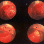
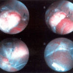
Falcons: The following images were obtained by endoscopy of falcons with aspergillosis.A,B Thoracic airsac (T) with prominent blood vessels and a dead serratospiculum worm (W). The presence of these lung worms makes the airsac look milky. D Normal ovary with developing follicles.

-
Falcons: The following images were obtained by endoscopy of falcons with aspergillosis.B,D Aspergillus lesions (A) over a swollen liver

-
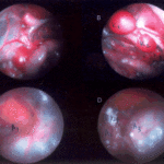
Falcons: The following images were obtained by endoscopy of falcons with aspergillosis.B Cranial, middle, caudal lobes (K1,K2,K3) of the left kidney, all the lobes show slight nephromegaly.C Yellow aspergillus colony (A1), lying adjacent to the lung.D White aspergillus colonies (A2,A3,A4).
-
Falcons: The following images were obtained by endoscopy of falcons with aspergillosis.C Cranial pole of left kidney (K) -mildly inflamed.D Ovary ( F) with developing follicles.
-
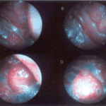
The following images were obtained by endoscopy of falcons with aspergillosis.A and B Lung Worm (S) over liver (Li) (serratospiculum seurati)C and D Aspergilloma (A) and prominent blood vessels on the caudal thoracic air sac (T).
