Date: 26 November 2013
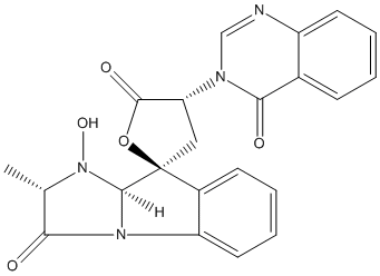
Secondary metabolites, structure diagram: Trivial name – tryptoquivaline E
Copyright: n/a
Notes:
Species: A. fumigatusSystematic name: Spiro[furan-2(5H),9′-[9H]imidazo[1,2-a]indole]-3′,5(2’H)-dione, 1′,3,4,9’a-tetrahydro-1′-hydroxy-2′-methyl-4-(4-oxo-3(4H)-quinazolinyl)-, (2S,2’S,4R,9’aS)-Molecular formulae: C22H18N4O5Molecular weight: 418.402Chemical abstracts number: 61897-87-8Selected references: Yamazaki, Mikio; Fujimoto, Haruhiro; Okuyama, Emi (Res. Inst. Chemobiodynamics, Chiba Univ., Chiba, Japan). Tetrahedron Lett., (33), 2861-4 (English) 1976.SECONDARY METABOLITES MYCOTOXINS PRODUCED BY FUNGI COLONIZING CEREAL GRAIN IN STORAGE STRUCTURE AND PROPERTIES GOLINSKI P CHELKOWSKI, J. (ED.). DEVELOPMENTS IN FOOD SCIENCE, VOL. 26. CEREAL GRAIN: MYCOTOXINS, FUNGI AND QUALITY IN DRYING AND STORAGE. XXII+607P. ELSEVIER SCIENCE PUBLISHERS B.V.: AMSTERDAM, NETHERLANDS; (DIST. IN THE USA AND CANADA BY ELSEVIER SCIENCE PUBLISHING CO., INC.: NEW YORK, NEW YORK, USA). ILLUS. MAPS. ISBN 0-444-88554-4.; 0 (0). 1991. 355-403.
Images library
-
Title
Legend
-
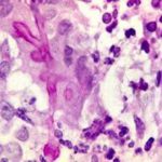
BAL specimen showing hyaline, septate hyphae consistent with Aspergillus, stained with Blankophor
-
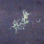
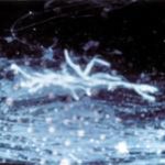
Mucous plug examined by light microscopy with KOH, showing a network of hyaline branching hyphae typical of Aspergillus, from a patient with ABPA.
-
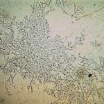
Corneal scraping stained with lactophenol cotton blue showing beaded septate hyphae not typical of either Fusarium spp or Aspergillus spp, being more consistent with a dematiceous (ie brown coloured) fungus
-
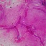
Corneal scrape with lactophenol cotton blue shows separate hyphae with Fusarium spp or Aspergillus spp.
-
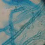
A filamentous fungus in the CSF of a patient with meningitis that grew Candida albicans in culture subsequently.
-
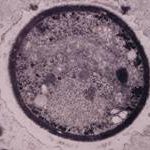
Transmission electron micrograph of a C. neoformans cell seen in CSF in an AIDS patients with remarkably little capsule present. These cells may be mistaken for lymphocytes.
-
India ink preparation of CSF showing multiple yeasts with large capsules, and narrow buds to smaller daughter cells, typical of C. neoformans
