Date: 26 November 2013
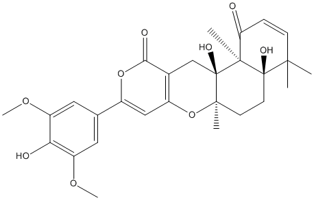
Secondary metabolites, structure diagram: Trivial name – territrem C
Copyright: n/a
Notes:
Species: A. terreusSystematic name: 4H,11H-Naphtho(2,1-b)pyrano(3,4-e)pyran-1,11(5H)-dione, 4a,6,6a,12,12a,12b-hexahydro-4a,12a-dihydroxy-9-(4-hydroxy-3,5-dimethoxyphenyl)-4,4,6a,12b-tetramethyl-, (4aR-(4a-alpha,6a-beta,12a-alpha,12b-beta))-Molecular formulae: C28H32O9Molecular weight: 512.548Chemical abstracts number: 89020-33-7Selected references: Ling KH, Yang CK, Peng FT. Territrems, tremorgenic mycotoxins of Aspergillus terreus. Appl Environ Microbiol. 1979 Mar;37(3):355-7. Ling KH, Liou HH, Yang CM, Yang CK. Related Articles, Links Isolation, chemical structure, acute toxicity, and some physicochemical properties of territrem C from Aspergillus terreus. Appl Environ Microbiol. 1984 Jan;47(1):98-100. Chen JW, Ling KH. Territrems: Naturally Occurring Specific Irreversible Inhibitors of Acetylcholinesterase. J Biomed Sci. 1996 Jan;3(1):54-58.Toxicity: mouse LD50 intraperitoneal 6280ug/kg (6.28mg/kg) Applied and Environmental Microbiology. Vol. 47, Pg. 98, 1984.
Images library
-
Title
Legend
-
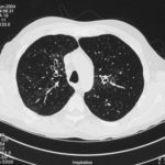
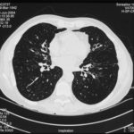
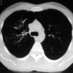
High resolution CT scan images with reconstruction of 1mm thick slices at approximately 10mm increments. The scan shows moderately severe multi-lobar cylindrical and varicose bronchiectasis predominantly centrally and in the upper lungs. There is no mucus plugging seen.
The features are in keeping with allergic bronchopulmonary aspergillosis
 ,
,  ,
, 
-
pt.SB – 6/10/98 – bronchocentric granulomatosis. CT scan showing multiple small nodules of variable size in both lung fields, apparently close to the vascular bundles.

-
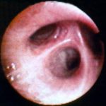
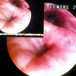
Bronchial oedema.Remarkably oedematous bronchial mucosa, as seen in ABPA.
-
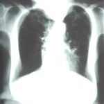
An example of longstanding allergic bronchopulmonary aspergillosis in a patient who has been steroid dependent for over 15 years showing remarkable kyphoscoliosis and honey combing and fibrosis of both lungs.
-
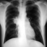
Recurrent pulmonary shadows 1. 6 Jan 1988 – chest radiograph showing right hilar enlargement, consistent with ABPA.
Recurrent pulmonary shadows 1. 3 Feb 1989 – chest radiograph showing right upper-lobe consolidation and contraction consistent with obstruction of RUL bronchus, in ABPA.
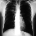
Clearing of pulmonary shadows 3, pt BJ. 5 April 1989 – resolution of shadows seen in February, with a course of corticosteroids.
Recurrence of pulmonary shadows 4, pt BJ. 2 September 1989 – recurrence of pulmonary shadows with an exacerbation of ABPA.
Central bronchiectasis, pt BJ. CT scan of thorax October 1989 showing central bronchiectasis, characteristic of ABPA (and cystic fibrosis).
 ,
,  ,
,  ,
,  ,
, 
-
A typical example of a wet mount of a sputum sample from a patient with allergic bronchopulmonary aspergillosis.


