Date: 26 November 2013
Secondary metabolites, structure diagram: Trivial name – sphingofungin C
Copyright: n/a
Notes:
Species: A. fumigatusSystematic name: 6-Eicosenoic acid, 5-(acetyloxy)-2-amino-3,4,14-trihydroxy-Molecular formulae: C22H41NO7Molecular weight: 431.563Chemical abstracts number: 121025-46-5Selected references: VanMiddlesworth F, Giacobbe RA, Lopez M, Garrity G, Bland JA, Bartizal K, Fromtling RA, Polishook J, Zweerink M, Edison AM, et al. J Antibiot (Tokyo). 1992 Jun;45(6):861-7. Sphingofungins A, B, C, and D; a new family of antifungal agents. I. Fermentation, isolation, and biological activity.Zweerink MM, Edison AM, Wells GB, Pinto W, Lester RL. Characterization of a novel, potent, and specific inhibitor of serine palmitoyltransferase. J Biol Chem. 1992 Dec 15;267(35):25032-8. FUMONISINS POWELL RG ; PLATTNER RD PELLETIER, S. W. (ED.). ALKALOIDS: CHEMICAL AND BIOLOGICAL PERSPECTIVES, VOL. 9. XV+286P. PERGAMON PRESS LTD: OXFORD, ENGLAND, UK; TARRYTOWN, NEW YORK, USA. ISBN 0-08-042089-3.; 0 (0). 1995. 247-278.
Images library
-
Title
Legend
-
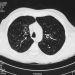
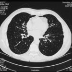
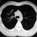
High resolution CT scan images with reconstruction of 1mm thick slices at approximately 10mm increments. The scan shows moderately severe multi-lobar cylindrical and varicose bronchiectasis predominantly centrally and in the upper lungs. There is no mucus plugging seen.
The features are in keeping with allergic bronchopulmonary aspergillosis
 ,
,  ,
, 
-
pt.SB – 6/10/98 – bronchocentric granulomatosis. CT scan showing multiple small nodules of variable size in both lung fields, apparently close to the vascular bundles.
-
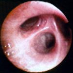
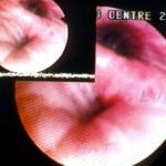
Bronchial oedema.Remarkably oedematous bronchial mucosa, as seen in ABPA.
-
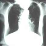
An example of longstanding allergic bronchopulmonary aspergillosis in a patient who has been steroid dependent for over 15 years showing remarkable kyphoscoliosis and honey combing and fibrosis of both lungs.
-
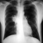
Recurrent pulmonary shadows 1. 6 Jan 1988 – chest radiograph showing right hilar enlargement, consistent with ABPA.
Recurrent pulmonary shadows 1. 3 Feb 1989 – chest radiograph showing right upper-lobe consolidation and contraction consistent with obstruction of RUL bronchus, in ABPA.
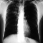
Clearing of pulmonary shadows 3, pt BJ. 5 April 1989 – resolution of shadows seen in February, with a course of corticosteroids.
Recurrence of pulmonary shadows 4, pt BJ. 2 September 1989 – recurrence of pulmonary shadows with an exacerbation of ABPA.
Central bronchiectasis, pt BJ. CT scan of thorax October 1989 showing central bronchiectasis, characteristic of ABPA (and cystic fibrosis).
 ,
,  ,
,  ,
,  ,
, 
-
A typical example of a wet mount of a sputum sample from a patient with allergic bronchopulmonary aspergillosis.


