Date: 26 November 2013
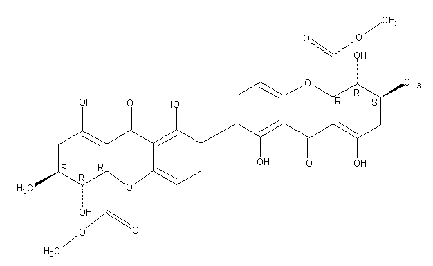
Secondary metabolites, structure diagram: Trivial name – secalonic acid D
Copyright: n/a
Notes:
Species: A. aculeatus, A. ochraceus, A. uvarumSystematic name: [7,7′-Bi-4aH-xanthene]-4a,4’a-dicarboxylic acid, 2,2′,3,3′,4,4′,9,9′-octahydro-1,1′,4,4′,8,8′-hexahydroxy-3,3′-dimethyl-9,9′-dioxo-, dimethyl ester, (3S,3’S,4R,4’R,4aR,4’aR)-Molecular formulae: C32 H30 O14Molecular weight: 638.581Chemical abstracts number: 35287-69-5Selected references: Andersen, Raymond; Buechi, George; Kobbe, Brunhilde; Demain, Arnold L. (Dep. Chem., Massachusetts Inst. Technol., Cambridge, Mass., USA). J. Org. Chem., 42(2), 352-3 (English) 1977.Kurobane I, Vining LC, McInnes AG. J Antibiot (Tokyo). 1979 Dec;32(12):1256-66. Biosynthetic relationships among the secalonic acids. Isolation of emodin, endocrocin and secalonic acids from Pyrenochaeta terrestris and Aspergillus aculeatus.Toxicity: mouse LD50 intraperitoneal 26500ug/kg (26.5mg/kg) EFFECTS: VASCULAR: REGIONAL OR GENERAL ARTERIOLAR OR VENOUS DILATION LUNGS, THORAX, OR RESPIRATION: CHANGES IN PULMONARY VASCULAR RESISTANCE LUNGS, THORAX, OR RESPIRATION: OTHER CHANGES Applied and Environmental Microbiology. Vol. 39, Pg. 285, 1980. mouse LD50 intravenous 25mg/kg (25mg/kg) EFFECTS: BEHAVIORAL: CONVULSIONS OR EFFECT ON SEIZURE THRESHOLD BEHAVIORAL: FOOD INTAKE (ANIMAL) SKIN AND APPENDAGES (SKIN): HAIR: OTHER Journal of Toxicology and Environmental Health. Vol. 5, Pg. 1159, 1979.mouse LDLo oral 30mg/kg (30mg/kg) EFFECTS: SENSE ORGANS AND SPECIAL SENSES: OTHER CHANGES: OLFACTION LIVER: HEPATITIS (HEPATOCELLULAR NECROSIS), ZONAL LUNGS, THORAX, OR RESPIRATION: OTHER CHANGES Toxicology and Applied Pharmacology. Vol. 48, Pg. A14, 1979. rat LD50 oral 22mg/kg (22mg/kg) EFFECTS: SENSE ORGANS AND SPECIAL SENSES: OTHER CHANGES: OLFACTION LIVER: HEPATITIS (HEPATOCELLULAR NECROSIS), ZONAL LUNGS, THORAX, OR RESPIRATION: OTHER CHANGES Toxicology and Applied Pharmacology. Vol. 48, Pg.
Images library
-
Title
Legend
-
Image A
CT Scan 30/3/99
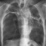
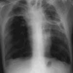
Showing extreme pleural thickening and 2 small cavities at apex of left lung.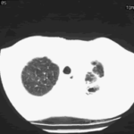
-
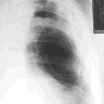
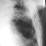
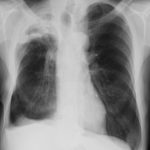
A 43 year old with smoking related emphysema was admitted to hospital with two separate episodes of haemoptysis. He had been in good health up to 1989, when he was diagnosed as having bilateral pulmonary tuberculosis. At that time a CT scan revealed a cavity in the left upper lobe (20.8cm2) with adjacent confluent infiltrates and pleural thickening. On bronchoscopic examination no abnormalities were noted and endobronchial biopsies did not reveal hyphae.
Over the next 4 years his condition deteriorated and a CT scan showed the left upper lobe cavity had increased to 40cm2. Itraconazole 400mg daily was prescribed. There was some clinical improvement on itraconazole but patient eventually deteriorated with breathlessness and with significant weight loss.
 ,
, 