Date: 26 November 2013
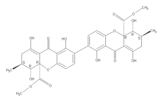
Secondary metabolites, structure diagram: Trivial name – secalonic acid D
Copyright: n/a
Notes:
Species: A. aculeatus, A. ochraceus, A. uvarumSystematic name: [7,7′-Bi-4aH-xanthene]-4a,4’a-dicarboxylic acid, 2,2′,3,3′,4,4′,9,9′-octahydro-1,1′,4,4′,8,8′-hexahydroxy-3,3′-dimethyl-9,9′-dioxo-, dimethyl ester, (3S,3’S,4R,4’R,4aR,4’aR)-Molecular formulae: C32 H30 O14Molecular weight: 638.581Chemical abstracts number: 35287-69-5Selected references: Andersen, Raymond; Buechi, George; Kobbe, Brunhilde; Demain, Arnold L. (Dep. Chem., Massachusetts Inst. Technol., Cambridge, Mass., USA). J. Org. Chem., 42(2), 352-3 (English) 1977.Kurobane I, Vining LC, McInnes AG. J Antibiot (Tokyo). 1979 Dec;32(12):1256-66. Biosynthetic relationships among the secalonic acids. Isolation of emodin, endocrocin and secalonic acids from Pyrenochaeta terrestris and Aspergillus aculeatus.Toxicity: mouse LD50 intraperitoneal 26500ug/kg (26.5mg/kg) EFFECTS: VASCULAR: REGIONAL OR GENERAL ARTERIOLAR OR VENOUS DILATION LUNGS, THORAX, OR RESPIRATION: CHANGES IN PULMONARY VASCULAR RESISTANCE LUNGS, THORAX, OR RESPIRATION: OTHER CHANGES Applied and Environmental Microbiology. Vol. 39, Pg. 285, 1980. mouse LD50 intravenous 25mg/kg (25mg/kg) EFFECTS: BEHAVIORAL: CONVULSIONS OR EFFECT ON SEIZURE THRESHOLD BEHAVIORAL: FOOD INTAKE (ANIMAL) SKIN AND APPENDAGES (SKIN): HAIR: OTHER Journal of Toxicology and Environmental Health. Vol. 5, Pg. 1159, 1979.mouse LDLo oral 30mg/kg (30mg/kg) EFFECTS: SENSE ORGANS AND SPECIAL SENSES: OTHER CHANGES: OLFACTION LIVER: HEPATITIS (HEPATOCELLULAR NECROSIS), ZONAL LUNGS, THORAX, OR RESPIRATION: OTHER CHANGES Toxicology and Applied Pharmacology. Vol. 48, Pg. A14, 1979. rat LD50 oral 22mg/kg (22mg/kg) EFFECTS: SENSE ORGANS AND SPECIAL SENSES: OTHER CHANGES: OLFACTION LIVER: HEPATITIS (HEPATOCELLULAR NECROSIS), ZONAL LUNGS, THORAX, OR RESPIRATION: OTHER CHANGES Toxicology and Applied Pharmacology. Vol. 48, Pg.
Images library
-
Title
Legend
-
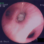
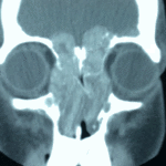
4 Total obstruction of the sinuses due to inflamed mucosa. (Patient 04)
-
1 Axial computed tomography (CT) scans of the frontal sinus.
A: due to the long lasting pressure of mucus, the bone of the anterior wall of frontal sinus is thinned out and elevated anteriorly, forming a bulge. B: same situation as depicted in fig A: the posterior bony wall of frontal sinus is thinned out and extremely elevated posteriorly towards the frontal lobe of the brain. As depicted on the scan, a thin bony layer covering the dura could be recognized intraoperatively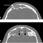
-
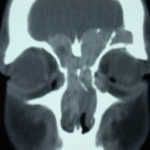
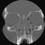
2 Same patient as 1 and 3, frontal CT
-
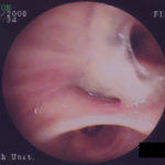
D. 6 months later, tenacious yellow secretions in L basal bronchial division
-
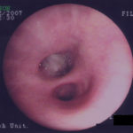
C. After suction the material was seen to extend distally – obstructing the right basal stem bronchus
-
B. After suction the material was seen to extend distally – obstructing the right basal stem bronchus
