Date: 26 November 2013
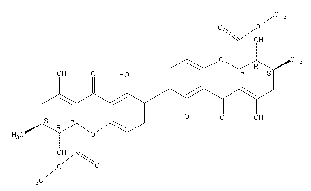
Secondary metabolites, structure diagram: Trivial name – secalonic acid D
Copyright: n/a
Notes:
Species: A. aculeatus, A. ochraceus, A. uvarumSystematic name: [7,7′-Bi-4aH-xanthene]-4a,4’a-dicarboxylic acid, 2,2′,3,3′,4,4′,9,9′-octahydro-1,1′,4,4′,8,8′-hexahydroxy-3,3′-dimethyl-9,9′-dioxo-, dimethyl ester, (3S,3’S,4R,4’R,4aR,4’aR)-Molecular formulae: C32 H30 O14Molecular weight: 638.581Chemical abstracts number: 35287-69-5Selected references: Andersen, Raymond; Buechi, George; Kobbe, Brunhilde; Demain, Arnold L. (Dep. Chem., Massachusetts Inst. Technol., Cambridge, Mass., USA). J. Org. Chem., 42(2), 352-3 (English) 1977.Kurobane I, Vining LC, McInnes AG. J Antibiot (Tokyo). 1979 Dec;32(12):1256-66. Biosynthetic relationships among the secalonic acids. Isolation of emodin, endocrocin and secalonic acids from Pyrenochaeta terrestris and Aspergillus aculeatus.Toxicity: mouse LD50 intraperitoneal 26500ug/kg (26.5mg/kg) EFFECTS: VASCULAR: REGIONAL OR GENERAL ARTERIOLAR OR VENOUS DILATION LUNGS, THORAX, OR RESPIRATION: CHANGES IN PULMONARY VASCULAR RESISTANCE LUNGS, THORAX, OR RESPIRATION: OTHER CHANGES Applied and Environmental Microbiology. Vol. 39, Pg. 285, 1980. mouse LD50 intravenous 25mg/kg (25mg/kg) EFFECTS: BEHAVIORAL: CONVULSIONS OR EFFECT ON SEIZURE THRESHOLD BEHAVIORAL: FOOD INTAKE (ANIMAL) SKIN AND APPENDAGES (SKIN): HAIR: OTHER Journal of Toxicology and Environmental Health. Vol. 5, Pg. 1159, 1979.mouse LDLo oral 30mg/kg (30mg/kg) EFFECTS: SENSE ORGANS AND SPECIAL SENSES: OTHER CHANGES: OLFACTION LIVER: HEPATITIS (HEPATOCELLULAR NECROSIS), ZONAL LUNGS, THORAX, OR RESPIRATION: OTHER CHANGES Toxicology and Applied Pharmacology. Vol. 48, Pg. A14, 1979. rat LD50 oral 22mg/kg (22mg/kg) EFFECTS: SENSE ORGANS AND SPECIAL SENSES: OTHER CHANGES: OLFACTION LIVER: HEPATITIS (HEPATOCELLULAR NECROSIS), ZONAL LUNGS, THORAX, OR RESPIRATION: OTHER CHANGES Toxicology and Applied Pharmacology. Vol. 48, Pg.
Images library
-
Title
Legend
-
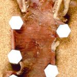
Pt FT. Autopsy appearance of the trachea, after the adherent pseudomembrane had been removed, revealing confluent ulceration superiorly with small green plaques of Aspergillus growth on the trachea inferiorly.
-
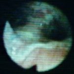
This view was obtained in a lung transplant recipient at bronchoscopy. Aspergillus fumigatus was grown from bronchial lavage but invasion was not demonstrated on bronchial biopsy. Symptoms improved with itraconazole therapy and abnormal appearances had resolved within 2 weeks.

-
Bronchoscopic view of Aspergillus tracheobronchitis. Bronchial lavage revealed hyphae in microscopy and cultures grew A.fumigatus. This man had received a lung transplant a few weeks before. Invasion of mucosa, but not cartilage, was demonstrated histologically. He responded rapidly to oral itraconazole.
-
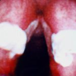
This view from indirect laryngoscopy illustrates bilateral lesions on the larynx that on biopsy were shown to be due to Aspergillus. This is a rare disease in non-immunocompromised patients.
-
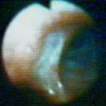
Bronchoscopic view of a deep bronchial ulcer in a lung transplant patient. Biopsies through the ulcer yielded cartilage with hyphae invading it. Fungal cultures of bronchial lavage grew Aspergillus fumigatus. He responded to oral itraconazole therapy.
-
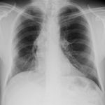
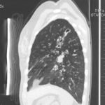
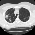
Patient had life threatening pneumonia, cavity formation was later observed. He later presented with a fungal ball. The aspergilloma was removed by surgical resection of the right upper lobe.
 ,
,  ,
,  ,
,  ,
,  ,
,  ,
, 