Date: 26 November 2013
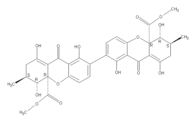
Secondary metabolites, structure diagram: Trivial name – secalonic acid D
Copyright: n/a
Notes:
Species: A. aculeatus, A. ochraceus, A. uvarumSystematic name: [7,7′-Bi-4aH-xanthene]-4a,4’a-dicarboxylic acid, 2,2′,3,3′,4,4′,9,9′-octahydro-1,1′,4,4′,8,8′-hexahydroxy-3,3′-dimethyl-9,9′-dioxo-, dimethyl ester, (3S,3’S,4R,4’R,4aR,4’aR)-Molecular formulae: C32 H30 O14Molecular weight: 638.581Chemical abstracts number: 35287-69-5Selected references: Andersen, Raymond; Buechi, George; Kobbe, Brunhilde; Demain, Arnold L. (Dep. Chem., Massachusetts Inst. Technol., Cambridge, Mass., USA). J. Org. Chem., 42(2), 352-3 (English) 1977.Kurobane I, Vining LC, McInnes AG. J Antibiot (Tokyo). 1979 Dec;32(12):1256-66. Biosynthetic relationships among the secalonic acids. Isolation of emodin, endocrocin and secalonic acids from Pyrenochaeta terrestris and Aspergillus aculeatus.Toxicity: mouse LD50 intraperitoneal 26500ug/kg (26.5mg/kg) EFFECTS: VASCULAR: REGIONAL OR GENERAL ARTERIOLAR OR VENOUS DILATION LUNGS, THORAX, OR RESPIRATION: CHANGES IN PULMONARY VASCULAR RESISTANCE LUNGS, THORAX, OR RESPIRATION: OTHER CHANGES Applied and Environmental Microbiology. Vol. 39, Pg. 285, 1980. mouse LD50 intravenous 25mg/kg (25mg/kg) EFFECTS: BEHAVIORAL: CONVULSIONS OR EFFECT ON SEIZURE THRESHOLD BEHAVIORAL: FOOD INTAKE (ANIMAL) SKIN AND APPENDAGES (SKIN): HAIR: OTHER Journal of Toxicology and Environmental Health. Vol. 5, Pg. 1159, 1979.mouse LDLo oral 30mg/kg (30mg/kg) EFFECTS: SENSE ORGANS AND SPECIAL SENSES: OTHER CHANGES: OLFACTION LIVER: HEPATITIS (HEPATOCELLULAR NECROSIS), ZONAL LUNGS, THORAX, OR RESPIRATION: OTHER CHANGES Toxicology and Applied Pharmacology. Vol. 48, Pg. A14, 1979. rat LD50 oral 22mg/kg (22mg/kg) EFFECTS: SENSE ORGANS AND SPECIAL SENSES: OTHER CHANGES: OLFACTION LIVER: HEPATITIS (HEPATOCELLULAR NECROSIS), ZONAL LUNGS, THORAX, OR RESPIRATION: OTHER CHANGES Toxicology and Applied Pharmacology. Vol. 48, Pg.
Images library
-
Title
Legend
-
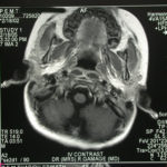
Image a. 3 yr old boy with CNS aspergillosis pt TS. MRI scan pre-amphotericin B
-
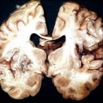
Section though unfixed brain showing large pale area of infarction deep in the parietal cortex, in which Aspergillus hyphae were seen histologically. The patient developed disseminated aspergillosis after a prolonged stay in intensive care after contracting severe community acquired pneumonia.
-
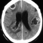
The woman had received a renal transplant several months prior to developing a stroke with reduced consciousness. The enhanced CT scan of her brain showed multiple ring-enhancing lesions bilaterally with little surrounding oedema. Biopsy confirmed invasive aspergillosis on histology and culture.
-
Further details
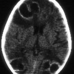
Image A. Multiple ring enhancing abscesses with substantial surrounding oedema was demonstrated. He had no focal neurological deficits. A needle aspiration confirmed the clinical impression of cerebral aspergillosis by culture and microscopy.
Image B. Resolution of cerebral aspergillosis, pt MN. Focal scars with some surrounding oedema are seen in the site of the prior abscesses.
 ,
, 
-
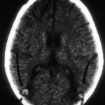
Contrast enhanced CT scan of the brain showing unequivocally 2 hypodense lesions, one in the left basal ganglia and one in the right occipital cortex. There is the possibility of another smaller left sided occiptal cortex. These lesions do not have the appearance of abscesses, but rather of ischaema.

-
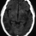
Unenhanced CT scan of the brain in an allogeneic bone marrow transplant recipient demonstrating a large, variably hypodense lesion in the area of the left basal ganglia and possible additional lesions in the posterior parietal and/or occipital cortex.
-
A. versicolor by microscopy showing very long thin conidiophores.

-
Pigmentation of Aspergillus versicolor colonies ranged from pale green to greenish-beige, pink-green, dark green and brown. Reverse is usually reddish. The growth rate is usually slow. Cultured on Sabouraud dextrose agar with chloramphenicol.

-
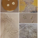
A Colonies on MEA after one week; B, C conidial heads with tip of conidiophire, x920; D conidial head, x 2330; E conidial heads x920
-
Cultures were grown on malt extract agar. Image kindly provided by Niall Hamilton.
