Date: 26 November 2013
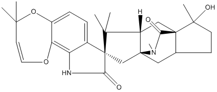
Secondary metabolites, structure diagram: Trivial name – paraherquamide
Copyright: n/a
Notes:
Species: A. silvaticusSystematic name: Spiro[4H,8H-[1,4]dioxepino[2,3-g]indole-8,7′(8’H)-[5H,6H-5a,9a](iminomethano)[1H]cyclopent[f]indolizine]-9,10′(10H)-dione, 2′,3′,8’a,9′-tetrahydro-1′-hydroxy-1′,4,4,8′,8′,11′-hexamethyl-, (1’R,5’aS,7’R,8’aS,9’aR)-Molecular formulae: C28H35N3O5Molecular weight: 493Chemical abstracts number: 77392-58-6Selected references: Yamazaki, M.; Fujimoto, H.; Okuyama, E.; Ohta, Y. (Res. Inst. Chemobiodyn., Chiba Univ., Chiba 280, Japan). Maikotokishin (Tokyo), 10, 27-8 (Japanese) 1980.
Images library
-
Title
Legend
-
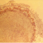
The periphery of the fungus ball is deeply eosinophilic because of the deposition of Splendore-Hoeppli material.
-
Single fungal ball, moving. Radiographic appearance of a fungus ball, showing movement as the patient’s position changes.
-
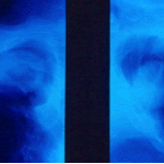
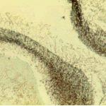
Oxalate crystals in the cavity wall surrounding an Aspergillus niger fungus ball (H&E, dark field, x 25).
-
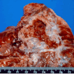
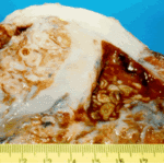
Aspergilloma patient. Gross pathology appearance of a fungus ball.
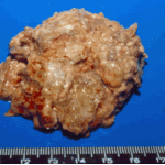
-
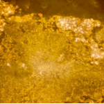
Conidiophores of Aspergillus fumigatus in the mass of the fungal ball surrounded by mycelia (H&E, x 400).
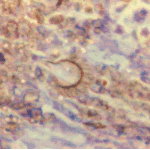
-
Aspergillus niger fungal ball. Calcium oxalate crystals in Aspergillus niger fungal ball. Also shown are darkly pigmented, rough-walled conidia associated with Aspergillus niger infection.
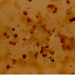
-
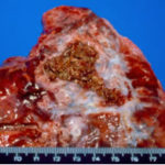
Aspergillus niger fungus ball within an old tuberculous cavern. This patient had diabetes, a disease commonly associated with A. niger infection.
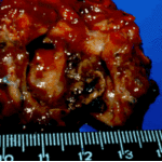

 ,
, 
 ,
, 