Date: 26 November 2013
Secondary metabolites, structure diagram: Trivial name – malformins
Copyright: n/a
Notes:
Species: A. nigerSystematic name: Cyclo(D-cysteinyl-D-cysteinyl-L-valyl-D-leucyl-L-leucyl), cyclic (1®2)-disulfideMalformin AMolecular formulae: C23H39N5O5S2Molecular weight: 529.718Chemical abstracts number: 3022-92-2Selected references: Kim KW, Sugawara F, Yoshida S, Murofushi N, Takahashi N, Curtis RW. Structure of malformin B, a phytotoxic metabolite produced by Aspergillus niger. Biosci Biotechnol Biochem. 1993 May;57(5):787-91.Toxicity: mouse LD50 intraperitoneal 3100ug/kg (3.1mg/kg) EFFECTS: GASTROINTESTINAL: OTHER CHANGES. LIVER: OTHER CHANGES. KIDNEY, URETER, AND BLADDER: OTHER CHANGES Agricultural and Biological Chemistry. Vol. 39, Pg. 1325, 1975.
Images library
-
Title
Legend
-
BAL specimen showing hyaline, septate hyphae consistent with Aspergillus, stained with Blankophor
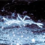
-
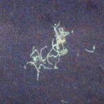
Mucous plug examined by light microscopy with KOH, showing a network of hyaline branching hyphae typical of Aspergillus, from a patient with ABPA.
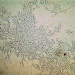
-
Corneal scraping stained with lactophenol cotton blue showing beaded septate hyphae not typical of either Fusarium spp or Aspergillus spp, being more consistent with a dematiceous (ie brown coloured) fungus
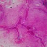
-
Corneal scrape with lactophenol cotton blue shows separate hyphae with Fusarium spp or Aspergillus spp.
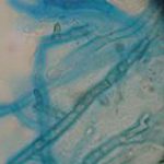
-
A filamentous fungus in the CSF of a patient with meningitis that grew Candida albicans in culture subsequently.
-
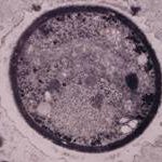
Transmission electron micrograph of a C. neoformans cell seen in CSF in an AIDS patients with remarkably little capsule present. These cells may be mistaken for lymphocytes.
-
India ink preparation of CSF showing multiple yeasts with large capsules, and narrow buds to smaller daughter cells, typical of C. neoformans
