Date: 26 November 2013
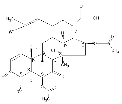
Secondary metabolites, structure diagram: Trivial name – helvolic acid
Copyright: n/a
Notes:
Species: A. fumigatiaffinis, A. fumigatus, A. novofumigatusSystematic name: 29-Nordammara-1,17(20),24-trien-21-oic acid, 6,16-bis(acetyloxy)-3,7-dioxo-, (4a,6b,8a,9b,13a,14b,16b,17Z)- (9CI)Helvolic acid (6CI, 7CI); (Z)-6b,16b-Dihydroxy-3,7-dioxo-29-nor-8a,9b,13a,14b-dammara-1,17(20),24-trien-21-oic acid diacetate; FumigacinMolecular formulae: C33H44O8Molecular weight: 568.698Chemical abstracts number: 29400-42-8Selected references: Waksman SA , et al. The production of two antibacterial substances, fumigacin and clavacin. Science 96: 202-203, 1942WILLIAMS TI.Biochem J. 1952 Jul;51(4):538-42. Some chemical properties of helvolic acid.OKUDA S, IWASAKI S, TSUDA K, SANO Y, HATA T, UDAGAWA S, NAKAYAMA Y, YAMAGUCHI H. THE STRUCTURE OF HELVOLIC ACID.Chem Pharm Bull (Tokyo). 1964 Jan;12:121-4. Amitani R, Taylor G, Elezis EN, Llewellyn-Jones C, Mitchell J, Kuze F, Cole PJ, Wilson R. Purification and characterization of factors produced by Aspergillus fumigatus which affect human ciliated respiratory epithelium. Infect Immun. 1995 Sep;63(9):3266-71.Toxicity: mouse LD50 intraperitoneal 400mg/kg (400mg/kg) Antibiotics: Origin, Nature, and Properties, Korzyoski, T., et al., eds., Washington, DC, American Soc. for Microbiology, 1978Vol. 3, Pg. 1837, 1978. mouse LDLo intravenous 500mg/kg (500mg/kg) Antibiotics: Origin, Nature, and Properties, Korzyoski, T., et al., eds., Washington, DC, American Soc. for Microbiology, 1978Vol. 3, Pg. 1837, 1978.
Images library
-
Title
Legend
-
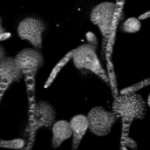
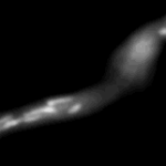
Mitochondria organisation: GFP fluorescence micrographs showing mitochondrial organisation in an A.nidulans strain with GFP mitochondria, grown at 25°C in minimal media.
-
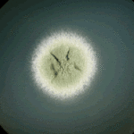
Colony morphology of A.nidulans SRF200 after two days at 37°C
-
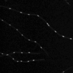
Aspergillus nidulans. Cell nuclei-Ds red. DsRed fluorescence micrographs showing nuclear distribution in an A.nidulans germling with dsRed stained nuclei

-
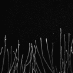
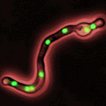
Cell Biology – Aspergillus nidulans. Cell nuclei-GFP. Nuclear distribution: GFP fluorescence mirographs showing fungal cell morphology and nuclear distribution in A.nidulans. GFP stained nuclei,grown at 25°C in minimal media O/N
-
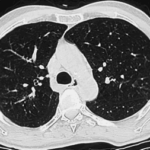
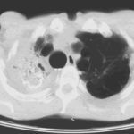
High resolution CT scan of chest.CT scan demonstrating remarkable bronchial wall thickening of the right main bronchus and main branches, in context of longstanding ABPA


