Date: 26 November 2013
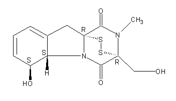
Secondary metabolites, structure diagram: Trivial name – gliotoxin
Copyright: n/a
Notes:
Species: A. flavus, A. fumigatus, A. niger, A. terreus, Eurotium chevalieri, Neosartorya pseudofischeriSystematic name: 10H-3,10a-Epidithiopyrazino[1,2-a]indole-1,4-dione, 2,3,5a,6-tetrahydro-6-hydroxy-3-(hydroxymethyl)-2-methyl-, (3R,5aS,6S,10aR)-Molecular formulae: C13H14N2O4S2Molecular weight: 326.393Chemical abstracts number: 67-99-2Selected references: Larsen TO, Smedsgaard J, Nielsen KF, Hansen MA, Samson RA, Frisvad JC. Production of mycotoxins by Aspergillus lentulus and other medically important and closely related species in section Fumigati. Med Mycol. 2007 May;45(3):225-32. Belkacemi, L.; Barton, R. C.; Hopwood, V.; Evans, E. G. V. (CORPORATE SOURCE PHLS Mycology Reference Laboratory, Department of Microbiology, University of Leeds, Leeds, UK). SOURCE Med. Mycol., 37(4), 227-233 (English) 1999 Blackwell Science Ltd. Lewis RE, Wiederhold NP, Lionakis MS, Prince RA, Kontoyiannis DP.J Clin Microbiol. 2005 Dec;43(12):6120-2. Frequency and species distribution of gliotoxin-producing Aspergillus isolates recovered from patients at a tertiary-care cancer center.Toxicity: Gliotoxin posseses a spectrum of biological activities including antibacterial and antiviral activities, and it is also a potent immunomodulating agent. Gliotoxin is also an inducer of apoptotic cell death in a number of cell types, and it has been found to be associated with some diseases attributed directly or indirectly to fungal infections. It is a secondary metabolite produced by a number of Aspergillus and Penicillium species.It is a potent immunosuppressive metabolite and brings about apoptosis in cells. Because of its effects on the immune system it may have a place in transplant surgery. There is limited evidence for its occurrence in moulded cereals. A. fumigatus is a potent pathogen which can colonise the lungs and other body tissues after ingestion of spores. There is some limited evidence that gliotoxin may be formed in situ in such circumstances. hamster LDLo oral 25mg/kg (25mg/kg) Veterinary and Human Toxicology. Vol. 32(Suppl), Pg. 63, 1990.mouse LD50 intraperitoneal 32mg/kg (32mg/kg) Chemotherapia. Vol. 10, Pg. 12, 1965. mouse LD50 intravenous 7800ug/kg (7.8mg/kg) Chemotherapia. Vol. 10, Pg. 12, 1965. mouse LD50 oral 67mg/kg (67mg/kg) Chemotherapia. Vol. 10, Pg. 12, 1965. mouse LD50 subcutaneous 25mg/kg (25mg/kg) Chemotherapia. Vol. 10, Pg. 12, 1965. rabbit LDLo intravenous 45mg/kg (45mg/kg) VASCULAR: BP LOWERING NOT CHARACTERIZED IN AUTONOMIC SECTION. GASTROINTESTINAL: HYPERMOTILITY, DIARRHEA Journal of the American Chemical Society. Vol. 65, Pg. 2005, 1943. rat LDLo intravenous 45mg/kg (45mg/kg) Veterinary and Human Toxicology. Vol. 32(Suppl), Pg. 63, 1990.rat LDLo unreported 50mg/kg (50mg/kg) BEHAVIORAL: ALTERED SLEEP TIME (INCLUDING CHANGE IN RIGHTING REFLEX) Journal of the American Chemical Society. Vol. 65, Pg. 2005, 1943.
Images library
-
Title
Legend
-
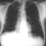
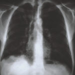
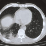
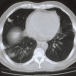
5 sputum samples were positive for A. niger, a bronchoscopy was normal and culture and microscopy were negative. Rx voriconazole, 200mg bd was given. An excellent clinical response was seen.
 ,
,  ,
,  ,
,  ,
, 
-
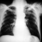
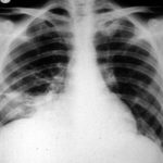
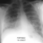
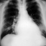
The patient was a 37-year old man in whom P.carinii pneumonia developed in August 1987, followed by esophageal candidiasis and upper gastrointestinal bleeding in September. Chronic perineal herpes led to the formation of rectourethral fistula and multiple episodes of urosepsis, for which he was given long-term ciprofloxacin therapy to suppress bacterial colonization of the bladder. He discontinued heavy alcohol use in September 1987 and smoked marijuana occasionally.On April 23 1989, the patient was admitted to the hospital with a two-month history of increasing dry cough with shortness of breath. He reported transient fever (temperature to 41°C). He was admitted with leukopenia, with his neutrophil count falling to 16 x 106/l on the second hospital day. A chest film showed bilateral fluffy lower-lobe infiltrates (this image). Zidovudine was discontinued. The patient had a rapidly downhill course despite intravenous treatment with trimethioprim-sulfamethoxazole. A bronchoscopy on the sixth hospital day revealed what appeared to be a foreign body in the left lower-lobe bronchus. It was removed, together with much necrotic, mucoid debris. On microscopic examination, the “foreign body” was necrotic, containing large numbers of hyphae and conidia in a manner typical of an aspegilloma or fungal cast. The culture grew A.fumigatus.
Clinical and radiologic improvement followed bronchoscopy, and itraconazole therapy was begun because of the concern about invasive aspergillosis in the setting of marked neutropenia. The patient tolerated the medication well at a dose of 200 mg twice daily, and the chest film became normal over the subsequent six weeks, after which itraconazole was discontinued. A sputum specimen cultured for fungus four weeks after the start of therapy was negative. After the initial improvement with itraconazole, the patient had recurrent urosepsis, associated with dehydration and marked confusion. Nine weeks after the discontinuation of itraconazole, he died of progressive dementia complicated by recurrent pneumonia and sepsis. There was no postmortem examination.
This patient was described (pt 11) and this chest radiograph reproduced in Denning DW, Follansbee S, Scolaro M, Norris S, Edelstein D, Stevens DA. Pulmonary Aspergillosis in the Acquired Immunodeficiency Syndrome. N Engl J Med 1991; 324: 654-662.
 ,
, 
-
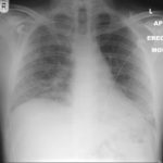
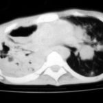
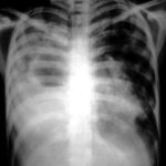
Image A. This 25 year old woman was previously well and presented with a pneumonia of uncertain aetiology. She has infiltrates in right upper-lobe and left middle and lower zones. The diagnosis was later made of chronic invasive pulmonary aspergillosis by bronchoscopy . Subsequently she was diagnosed with adult-onset chronic granulomatous disease with neutrophil function assays.
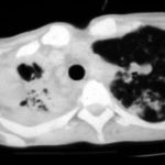
Image B. CT scan of the thorax just below the carina, showing almost complete opacification of the right lung and marked nodular shadowing around the hilum of the left lung.
Image C. Progression of pulmonary infiltrates are seen seven weeks later, despite administration of amphotericin B.
Image D. CT scan of the thorax above the carina showing near complete opacification of the right lung and multiple discrete nodular shadows in the left lung.
 ,
,  ,
,  ,
, 
-
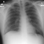
02/10/09 X -ray shows improvement in appearances from initial presentation with complete chest radiographic resolution by 07/11/2008
-
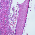
Subacute IPA in rhematoid nodules of the lung. in a patient with rheumatoid arthritis. Histology sections stained with H&E
