Date: 26 November 2013
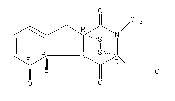
Secondary metabolites, structure diagram: Trivial name – gliotoxin
Copyright: n/a
Notes:
Species: A. flavus, A. fumigatus, A. niger, A. terreus, Eurotium chevalieri, Neosartorya pseudofischeriSystematic name: 10H-3,10a-Epidithiopyrazino[1,2-a]indole-1,4-dione, 2,3,5a,6-tetrahydro-6-hydroxy-3-(hydroxymethyl)-2-methyl-, (3R,5aS,6S,10aR)-Molecular formulae: C13H14N2O4S2Molecular weight: 326.393Chemical abstracts number: 67-99-2Selected references: Larsen TO, Smedsgaard J, Nielsen KF, Hansen MA, Samson RA, Frisvad JC. Production of mycotoxins by Aspergillus lentulus and other medically important and closely related species in section Fumigati. Med Mycol. 2007 May;45(3):225-32. Belkacemi, L.; Barton, R. C.; Hopwood, V.; Evans, E. G. V. (CORPORATE SOURCE PHLS Mycology Reference Laboratory, Department of Microbiology, University of Leeds, Leeds, UK). SOURCE Med. Mycol., 37(4), 227-233 (English) 1999 Blackwell Science Ltd. Lewis RE, Wiederhold NP, Lionakis MS, Prince RA, Kontoyiannis DP.J Clin Microbiol. 2005 Dec;43(12):6120-2. Frequency and species distribution of gliotoxin-producing Aspergillus isolates recovered from patients at a tertiary-care cancer center.Toxicity: Gliotoxin posseses a spectrum of biological activities including antibacterial and antiviral activities, and it is also a potent immunomodulating agent. Gliotoxin is also an inducer of apoptotic cell death in a number of cell types, and it has been found to be associated with some diseases attributed directly or indirectly to fungal infections. It is a secondary metabolite produced by a number of Aspergillus and Penicillium species.It is a potent immunosuppressive metabolite and brings about apoptosis in cells. Because of its effects on the immune system it may have a place in transplant surgery. There is limited evidence for its occurrence in moulded cereals. A. fumigatus is a potent pathogen which can colonise the lungs and other body tissues after ingestion of spores. There is some limited evidence that gliotoxin may be formed in situ in such circumstances. hamster LDLo oral 25mg/kg (25mg/kg) Veterinary and Human Toxicology. Vol. 32(Suppl), Pg. 63, 1990.mouse LD50 intraperitoneal 32mg/kg (32mg/kg) Chemotherapia. Vol. 10, Pg. 12, 1965. mouse LD50 intravenous 7800ug/kg (7.8mg/kg) Chemotherapia. Vol. 10, Pg. 12, 1965. mouse LD50 oral 67mg/kg (67mg/kg) Chemotherapia. Vol. 10, Pg. 12, 1965. mouse LD50 subcutaneous 25mg/kg (25mg/kg) Chemotherapia. Vol. 10, Pg. 12, 1965. rabbit LDLo intravenous 45mg/kg (45mg/kg) VASCULAR: BP LOWERING NOT CHARACTERIZED IN AUTONOMIC SECTION. GASTROINTESTINAL: HYPERMOTILITY, DIARRHEA Journal of the American Chemical Society. Vol. 65, Pg. 2005, 1943. rat LDLo intravenous 45mg/kg (45mg/kg) Veterinary and Human Toxicology. Vol. 32(Suppl), Pg. 63, 1990.rat LDLo unreported 50mg/kg (50mg/kg) BEHAVIORAL: ALTERED SLEEP TIME (INCLUDING CHANGE IN RIGHTING REFLEX) Journal of the American Chemical Society. Vol. 65, Pg. 2005, 1943.
Images library
-
Title
Legend
-
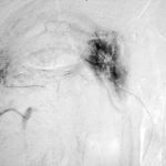
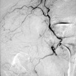
Late (venous) phase angiogram of a right intercostal artery showing persistence of vascular blush and further filling of a branch of the pulmonary artery.
-
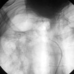
Catheter tip in a right posterior intercostal artery on screening film.
-
Angiogram of a right bronchial artery on subtraction film in the early arterial phase showing filling hypervascular circulation superiorly and communications with a pulmonary arterial radical.
-
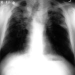
This patient with severe pulmonary sarcoidosis has bilateral aspergillomas. A rim of air is visible around parts of the aspergillomas on both sides. This patient was recruited into the NIAID Mycoses Study Group multicentre study of the treatment of invasive pulmonary aspergillosis with itraconazole but not analysed because invasive disease was not demonstrated. Denning DW, Lee JY, Hostetler JS, Pappas P, Kauffman CA, Dewsnup DH, Galgiani JN, Graybill JR, Sugar AM, Catanzaro A, Gallis H, Perfect JR, Dockery B, Dismukes WE, Stevens DA, NIAID Mycoses Study Group multicenter trial of oral itraconazole therapy of invasive aspergillosis. Am J Med 1994; 97: 135-144
-
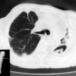
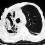
Extensive pleural thickening is demonstrated at the left apex on this CT scan of a woman who had previously had tuberculosis and whose large cavity gradually became obliterated by pleural thickening. An aspergilloma is demonstrable within the cavity
-
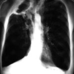
This chest radiograph (AMBER film) demonstrates the typical extensive pleural thickening at the right apex, seen in patients with aspergillomas. The cavity appears not to contain an aspergilloma but on CT scan had some ‘debris’ and Aspergillus antibiotics (precipitins) were strongly positive. The differential diagnosis lies between an aspergilloma and chronic invasive pulmonary aspergillosis. The extensive pleural thickening is heavily in favour of an aspergilloma, even without a well demonstrated fungal ball in the cavity.
-
Image C. Another example of a severe apical aspergilloma with remarkably little pleural thickening on plain chest radiograph (AMBER film). Severe distortion of the trachea is demonstrated.
 ,
,  ,
, 
-
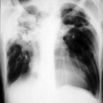
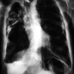
Right apical aspergilloma, patient WC. Plain chest radiograph of patient with right apical aspergilloma in an old, large tuberculous cavity. Severe haemoptysis and respiratory insufficiency together constituted the indications for embolisation which was done in one session over a 3 hour period (see images 1-6).
