Date: 26 November 2013
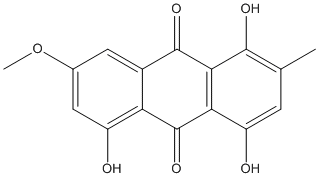
Secondary metabolites, structure diagram: trivial name – erythroglaucin
Copyright: n/a
Notes:
Species: A. chevalieriSystematic name: ANTHRAQUINONE, 7-METHOXY-2-METHYL-1,4,5-TRIHYDROXY- 1,4,5-Trihydroxy-7-methoxy-2-methylanthra-9,10-quinone 9,10-Anthracenedione, 1,4,5-trihydroxy-7-methoxy-2-methyl-Molecular formulae: C16H12O6Molecular weight: 300.263Chemical abstracts number: 476-57-3Selected references: Bachmann M, Blaser P, Luthy J, Schlatter C. J Environ Pathol Toxicol Oncol. 1992 Mar-Apr;11(2):49-52. Toxicity and mutagenicity of anthraquinones from Aspergillus chevalieri.
Images library
-
Title
Legend
-
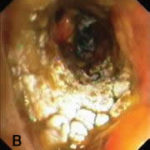
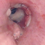
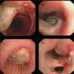
Bronchoscopic manifestations of Aspergillus tracheobronchitis. (a) Type I. Inflammatory infiltration, mucosa hyperaemia and plaques of pseudomembrane formation in the lumen without obvious airway occlusion. (b) Type II. Deep ulceration of the bronchial wall. (c) Type III. Significant airway occlusion by thick mucous plugs full of Aspergillus without definite deeper tissue invasion. (d) Type IV. Extensive tissue necrosis and pseudomembrane formation in the lumen with airway structures and severe airway occlusion (Wu 2010).
-
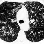
High resolution CT showing centrilobular nodular opacities and branching linear opacities (tree-in-bud appearance) (Al-Alawi 2007).
-
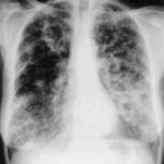
Chest X-ray showing poorly defined bilateral nodular opacities (Al-Alawi 2007).
-
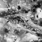
Gross pathologic specimen from autopsy shows the bronchial lumen covered by multiple whitish endobronchial nodules (arrows) (Franquet 2002).
-
Invasive tracheobronchitis showing numerous nodules seen during bronchoscopy (Ronan D’Driscoll).

-
Pseudomembranous seen overlying the bronchial mucosa (Tasci 2006).
