Date: 26 November 2013
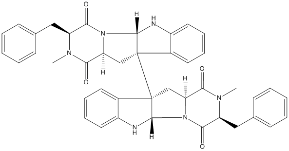
Secondary metabolites, structure diagram: Trivial name – ditryptophenoline
Copyright: n/a
Notes:
Species: A. flavusSystematic name: [10b,10’b-Bi-10bH-pyrazino[1′,2′:1,5]pyrrolo[2,3-b]indole]-1,1′,4,4′-tetrone, 2,2′,3,3′,5a,5’a,6,6′,11,11′,11a,11’a-dodecahydro-2,2′-dimethyl-3,3′-bis(phenylmethyl)-, (3S,3’S,5aS,5’aS,10bS,10’bS,11aS,11’aS)-Molecular formulae: C42H40N6O4Molecular weight: 692Chemical abstracts number: 64947-43-9Selected references: Springer, James P.; Buechi, George; Kobbe, Brunhilde; Demain, Arnold L.; Clardy, Jon (Dep. Chem., Massachusetts Inst. Technol., Cambridge, Mass., USA). Tetrahedron Lett., (28), 2403-6 (English) 1977.
Images library
-
Title
Legend
-
Further details
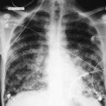
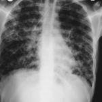
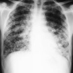
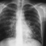
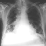
Image 1. The chest x-ray shows extensive bilateral nodular disease, most consistent with a fungal infection, or possibly tuberculosis. He was treated with a bucket face mask with 80% oxygen and voriconazole.
 ,
,  ,
,  ,
,  ,
,  ,
, 
-
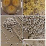
A Colonies on MEA +20 % sucrose after 2 weeks; B ascomata, x 40; C conidiophore of Aspergillus glaucus x 920;D conidiophore of Aspergillus glaucus x920 E. portion of ascoma with asci x 920. F ascospores x2330.
-
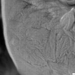
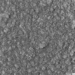
Scanning electron micrographs of A. fumigatus conidia of transformants rodB-02 (b). Size bar, 100 nm.

-
Scanning electron micrographs of A. fumigatus conidia of the wild-type G10 strain (a). Size bar, 100 nm.
-
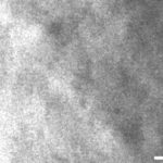
Scanning electron micrographs of A. fumigatus conidia of rodA rodB-26 (d).Size bar, 100 nm.
-
Scanning electron micrograph of an A.fumigatus conidium of rodA-47 (c), showing the hydrophobic rodlets covering the surface. Size bar, 100 nm.

 ,
, 
