Date: 26 November 2013
Secondary metabolites, structure diagram: Trivial name – Dehydroaustin
Copyright: n/a
Notes:
Species: A. niger, A. stellatus, A. ustus, Emericella dentataSystematic name: Spiro[1H,4H,8H-3a,7a-epoxy-6,11b-methano-3H-furo[3,4-e][3]benzoxocin-9(10H),3′(6’H)-[2H]pyran]-1,4,6′-trione, 7-(acetyloxy)-6,7,11,11a-tetrahydro-2′,2′,3,6,11a-pentamethyl-8,12-bis(methylene)-, (3R,3’R,3aR,6S,7R,7aR,11aR,11bS)-Molecular formulae: C27H30O9Molecular weight: 498Chemical abstracts number: 82893-35-4Selected references: Maebayashi, Yukio; Okuyama, Emi; Yamazaki, Mikio; Katsube, Yukiteru. Structure of ED-1 isolated from Emericella dentata. Chemical & Pharmaceutical Bulletin (1982), 30(5), 1911-12. Simpson, Thomas J.; Stenzel, Desmond J.; Bartlett, Alan J.; O’Brien, Eugene; Holker, John S. E. Studies on fungal metabolites. Part 3. Carbon-13 NMR spectral and structural studies on austin and new related meroterpenoids from Aspergillus ustus, Aspergillus variecolor, and Penicillium diversum. Journal of the Chemical Society, Perkin Transactions 1: Organic and Bio-Organic Chemistry (1972-1999) (1982), (11), 2687-92.
Images library
-
Title
Legend
-
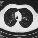
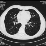
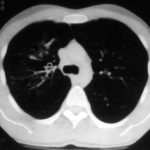
High resolution CT scan images with reconstruction of 1mm thick slices at approximately 10mm increments. The scan shows moderately severe multi-lobar cylindrical and varicose bronchiectasis predominantly centrally and in the upper lungs. There is no mucus plugging seen.
The features are in keeping with allergic bronchopulmonary aspergillosis
 ,
,  ,
, 
-
pt.SB – 6/10/98 – bronchocentric granulomatosis. CT scan showing multiple small nodules of variable size in both lung fields, apparently close to the vascular bundles.
-
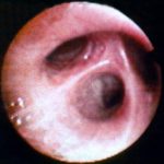
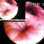
Bronchial oedema.Remarkably oedematous bronchial mucosa, as seen in ABPA.
-
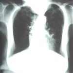
An example of longstanding allergic bronchopulmonary aspergillosis in a patient who has been steroid dependent for over 15 years showing remarkable kyphoscoliosis and honey combing and fibrosis of both lungs.
-
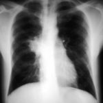
Recurrent pulmonary shadows 1. 6 Jan 1988 – chest radiograph showing right hilar enlargement, consistent with ABPA.
Recurrent pulmonary shadows 1. 3 Feb 1989 – chest radiograph showing right upper-lobe consolidation and contraction consistent with obstruction of RUL bronchus, in ABPA.
Clearing of pulmonary shadows 3, pt BJ. 5 April 1989 – resolution of shadows seen in February, with a course of corticosteroids.
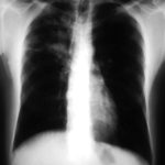
Recurrence of pulmonary shadows 4, pt BJ. 2 September 1989 – recurrence of pulmonary shadows with an exacerbation of ABPA.
Central bronchiectasis, pt BJ. CT scan of thorax October 1989 showing central bronchiectasis, characteristic of ABPA (and cystic fibrosis).
 ,
,  ,
,  ,
,  ,
, 
-
A typical example of a wet mount of a sputum sample from a patient with allergic bronchopulmonary aspergillosis.


