Date: 26 November 2013
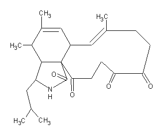
Secondary metabolites, structure diagram: Trivial name – aspochalasin A
Copyright: n/a
Notes:
Species: A. microcysticusSystematic name: 1H-Cycloundec[d]isoindole-1,15(2H)-dione, 3,3a,4,6a,9,10,11,12-octahydro-11,12-dihydroxy-4,5,8-trimethyl-3-(2-methylpropyl)-, (3S,3aR,4S,6aS,7E,11S,12S,13E,15aS)-Molecular formulae: C24H33NO4Molecular weight: 401Chemical abstracts number: 71968-02-0Selected references: Keller-Schierlein, Walter; Kupfer, Ernst (Org. Chem. Lab., ETH, Zurich CH-8092, Switz.). Helv. Chim. Acta, 62(5), 1501-24 (German) 1979.Toxicity Toxicity unknown
Images library
-
Title
Legend
-
Further details
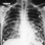
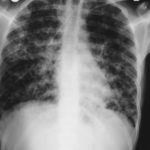
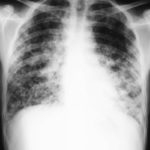
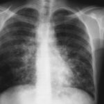
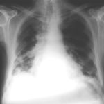
Image 1. The chest x-ray shows extensive bilateral nodular disease, most consistent with a fungal infection, or possibly tuberculosis. He was treated with a bucket face mask with 80% oxygen and voriconazole.
 ,
,  ,
,  ,
,  ,
,  ,
, 
-
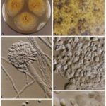
A Colonies on MEA +20 % sucrose after 2 weeks; B ascomata, x 40; C conidiophore of Aspergillus glaucus x 920;D conidiophore of Aspergillus glaucus x920 E. portion of ascoma with asci x 920. F ascospores x2330.
-
Scanning electron micrographs of A. fumigatus conidia of transformants rodB-02 (b). Size bar, 100 nm.

-
Scanning electron micrographs of A. fumigatus conidia of the wild-type G10 strain (a). Size bar, 100 nm.
-
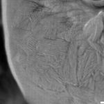
Scanning electron micrographs of A. fumigatus conidia of rodA rodB-26 (d).Size bar, 100 nm.
-
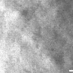
Scanning electron micrograph of an A.fumigatus conidium of rodA-47 (c), showing the hydrophobic rodlets covering the surface. Size bar, 100 nm.

 ,
, 
