Date: 26 November 2013
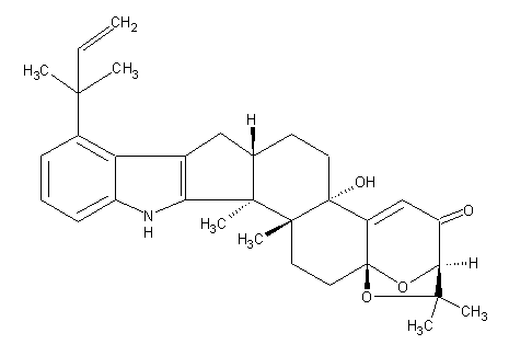
Secondary metabolites, structure diagram: Trivial name – aflatrem
Copyright: n/a
Notes:
Species: A. flavus, A. minisclerotigenesSystematic name: 4H-3,15a-Epoxy-1-benzoxepino[6′,7′:6,7]indeno[1,2-b]indol-4-one, 9-(1,1-dimethyl-2-propenyl)-2,3,5b,6,7,7a,8,13,13b,13c,14,15-dodecahydro-5b-hydroxy-2,2,13b,13c-tetramethyl-, (3R,5bS,7aS,13bS,13cR,15aS)-Molecular formulae: C32H39NO4Molecular weight: 501.67Chemical abstracts number: 70553-75-2Selected references: Cole, Richard J.; Dorner, Joe W.; Springer, James P.; Cox, Richard H. (Natl. Peanut Res. Lab., Sci. Educ. Adm., Dawson, GA 31742, USA). J. Agric. Food Chem., 29(2), 293-5 (English) 1981.Toxicity: The first tremorgenic mycotoxin to be isolated. An oral dose of 1 mg given to mice causes tremors, especially in response to attempts to move. The symptoms increase in severity after 1-2 h. but recovery usually occurs after 24-48 h.
Images library
-
Title
Legend
-
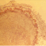
The periphery of the fungus ball is deeply eosinophilic because of the deposition of Splendore-Hoeppli material.
-
Single fungal ball, moving. Radiographic appearance of a fungus ball, showing movement as the patient’s position changes.
-
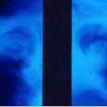
Oxalate crystals in the cavity wall surrounding an Aspergillus niger fungus ball (H&E, dark field, x 25).
-
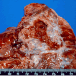
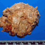
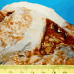
Aspergilloma patient. Gross pathology appearance of a fungus ball.
-
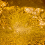
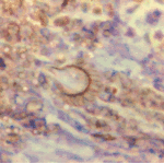
Conidiophores of Aspergillus fumigatus in the mass of the fungal ball surrounded by mycelia (H&E, x 400).
-
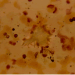
Aspergillus niger fungal ball. Calcium oxalate crystals in Aspergillus niger fungal ball. Also shown are darkly pigmented, rough-walled conidia associated with Aspergillus niger infection.
-
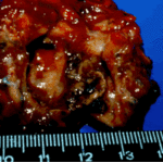
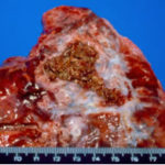
Aspergillus niger fungus ball within an old tuberculous cavern. This patient had diabetes, a disease commonly associated with A. niger infection.

 ,
, 
 ,
, 