Date: 26 November 2013
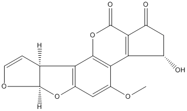
Secondary metabolites, structure diagram: Trivial name – aflatoxin Q1
Copyright: n/a
Notes:
Species: A. flavusSystematic name: Cyclopenta[c]furo[3′,2′:4,5]furo[2,3-h][1]benzopyran-1,11-dione, 2,3,6a,9a-tetrahydro-3-hydroxy-4-methoxy-, [3S-(3a,6aa,9aa)]-Molecular formulae: C17H12O7Molecular weight: 328Chemical abstracts number: 52819-96-2Selected references: Nakazato, Mitsuo; Morozumi, Satoshi; Saito, Kazuo; Fujinuma, Kenji; Nishima, Taichiro; Kasai, Nobuhiko (Dep. Food Hyg. and Nutr., Tokyo Metrop. Res. Lab. Public Health, Tokyo 169, Japan). Eisei Kagaku, 37(2), 107-16 (English) 1991.
Images library
-
Title
Legend
-
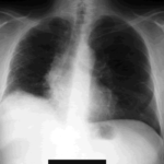
The chest x-ray shows a patient who had a left lung transplanted in May 2003 for cryptogenic fibrosing alveolitis, which was diagnosed post-transplant as sarcoidosis.
-
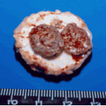
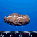
Gross pathology demonstrating the great pleural thickness and two cavities (upper lobe and superior segment of lower lobe) with fragments of fungal mass.

-
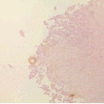
Histopathological appearance of a fungus ball. Note a conidial head resulting from fungal exposure to the air.
-
Histopathological appearance of a fungus ball caused by Scedosporium apiospermum. The presence of anneloconidia differentiates it from Aspergillus.

-
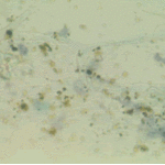
Chronic necrotising aspergillosis. Hyaline hyphal and calcium oxalate crystals obtained by needle aspirate biopsy from a diabetic patient with chronic necrotizing aspergillosis caused by Aspergillus niger (Papanicolaou, x 100).
-
Aspergillus niger fungus ball and acute oxalosis. Higher magnification of adjacent replicate section.
-
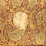
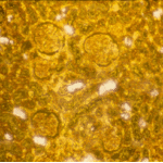
Oxalate crystals within renal tubuli (H&E, phase contrast, x 100). This patient developed acute oxalosis.
-
Lung surface. Fungus ball, severe parenchymal fibrosis and pleural thickening.


 ,
, 
 ,
,  ,
, 