Date: 26 November 2013
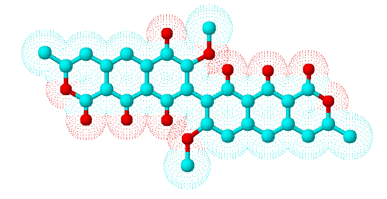
Secondary metabolites, 3D structure: Trivial name – viomellein
Copyright: n/a
Notes:
Species: A. melleus, A. ochraceusSystematic name: (8,8′-BI-1H-NAPHTHO(2,3-c)PYRAN)-1,1′,6,9-TETRONE, 3,3′,4,4′-TETRAHYDRO-7,7′-DIM (8,8′-Bi-1H-naphtho(2,3-c)pyran)-1,1′,6,9-tetrone, 3,3′,4,4′-tetrahydro-7,7′-dimethoxy-3,3′-dimethyl-9′,10,10′-trihydroxy-, (R-(R*,R*))-Molecular formulae: C30H24O11Molecular weight: 560.505Chemical abstracts number: 55625-78-0Selected references: Stack ME, Mislivec PB. Production of xanthomegnin and viomellein by isolates of Aspergillus ochraceus, Penicillium cyclopium, and Penicillium viridicatum. Appl Environ Microbiol. 1978 Oct;36(4):552-4. Filtenborg O, Frisvad JC, Svendsen JA. Simple screening method for molds producing intracellular mycotoxins in pure cultures. Appl Environ Microbiol. 1983 Feb;45(2):581-5. Robbers JE, Hong S, Tuite J, Carlton WW. Production of xanthomegnin and viomellein by species of Aspergillus correlated with mycotoxicosis produced in mice. Appl Environ Microbiol. 1978 Dec;36(6):819-23.Toxicity: Causes hepatic and renal damage in animals and associated with porcine nephropathy together with ochratoxins
Images library
-
Title
Legend
-
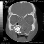
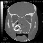
Patient MB X rays and CT scans. Chronic calcified maxillary sinusitis, patient had a palate defect.A. fumigatus cultured.
Images A&B Plain X rays antero-posterior and lateral, pre-operatively of Pt MB aged 76 who presented with unilateral nasal stuffiness and difficulty getting dentures fitted. She had hda these symptoms for many years. A large irregular calcified mass can be seen replacing the right maxillary sinus.
Images C D & E Coronal CT scan images of Pt MB showing a completely obstructed nasal cavity bilaterally and loss of internal nasal architecture. On the right side is large lamellar calcified lesion embedded in the extensive inflammatory material. Loss of bony margins is seen in numerous locations. This material was all removed surgically and showed mostly necrotic debris with Charcot-Leyden crystals and a few eosinophils and degenerate fungal hyphae. Aspergillus fumigatus was cultured from the material, especially infero-laterally on the right.
Image F Photograph through the mouth post-operatively showing the palate and a large defect in its right side. Through the defect can be seen the interior of the right maxillary sinus and nasal cavity with the inferior turbinate just visible.
 ,
,  ,
,  ,
,  ,
,  ,
, 
-
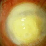
Aspergillus keratitis. Severe aspergillus infection with large area of corneal ulceration and deep stromal involvement

-
Sequence of images showing ocular surface change which unusually predisposed to severe fusarium keratitis in an elderly woman. Successful treatment involved full thickness corneal transplantation shown 2 weeks and then 2 years after surgery.

-
Sequence of images showing ocular surface change which unusually predisposed to severe fusarium keratitis in an elderly woman. Successful treatment involved full thickness corneal transplantation shown 2 weeks and then 2 years after surgery.
-
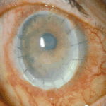
Sequence of images showing ocular surface change which unusually predisposed to severe fusarium keratitis in an elderly woman. Successful treatment involved full thickness corneal transplantation shown 2 weeks and then 2 years after surgery.
-
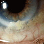
Sequence of images showing ocular surface change which unusually predisposed to severe fusarium keratitis in an elderly woman. Successful treatment involved full thickness corneal transplantation shown 2 weeks and then 2 years after surgery
-
Aspergillus keratitis. Shrunken eye as a consequence of this infection


