Date: 26 November 2013
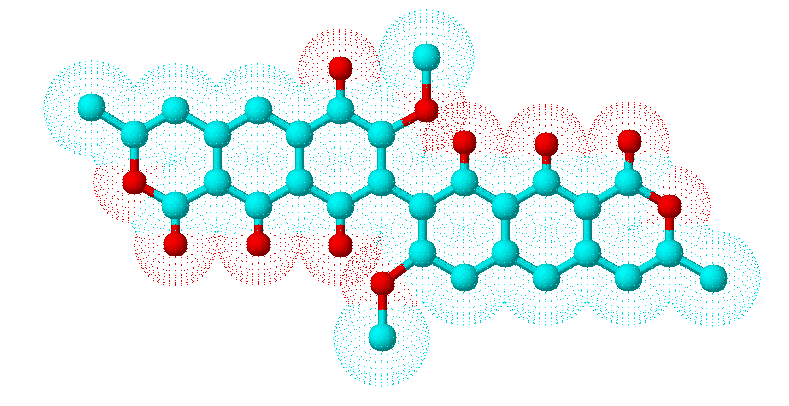
Secondary metabolites, 3D structure: Trivial name – viomellein
Copyright: n/a
Notes:
Species: A. melleus, A. ochraceusSystematic name: (8,8′-BI-1H-NAPHTHO(2,3-c)PYRAN)-1,1′,6,9-TETRONE, 3,3′,4,4′-TETRAHYDRO-7,7′-DIM (8,8′-Bi-1H-naphtho(2,3-c)pyran)-1,1′,6,9-tetrone, 3,3′,4,4′-tetrahydro-7,7′-dimethoxy-3,3′-dimethyl-9′,10,10′-trihydroxy-, (R-(R*,R*))-Molecular formulae: C30H24O11Molecular weight: 560.505Chemical abstracts number: 55625-78-0Selected references: Stack ME, Mislivec PB. Production of xanthomegnin and viomellein by isolates of Aspergillus ochraceus, Penicillium cyclopium, and Penicillium viridicatum. Appl Environ Microbiol. 1978 Oct;36(4):552-4. Filtenborg O, Frisvad JC, Svendsen JA. Simple screening method for molds producing intracellular mycotoxins in pure cultures. Appl Environ Microbiol. 1983 Feb;45(2):581-5. Robbers JE, Hong S, Tuite J, Carlton WW. Production of xanthomegnin and viomellein by species of Aspergillus correlated with mycotoxicosis produced in mice. Appl Environ Microbiol. 1978 Dec;36(6):819-23.Toxicity: Causes hepatic and renal damage in animals and associated with porcine nephropathy together with ochratoxins
Images library
-
Title
Legend
-
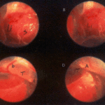
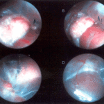
Falcons: The following images were obtained by endoscopy of falcons with aspergillosis.A,B Thoracic airsac (T) with prominent blood vessels and a dead serratospiculum worm (W). The presence of these lung worms makes the airsac look milky. D Normal ovary with developing follicles.

-
Falcons: The following images were obtained by endoscopy of falcons with aspergillosis.B,D Aspergillus lesions (A) over a swollen liver

-
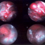
Falcons: The following images were obtained by endoscopy of falcons with aspergillosis.B Cranial, middle, caudal lobes (K1,K2,K3) of the left kidney, all the lobes show slight nephromegaly.C Yellow aspergillus colony (A1), lying adjacent to the lung.D White aspergillus colonies (A2,A3,A4).
-
Falcons: The following images were obtained by endoscopy of falcons with aspergillosis.C Cranial pole of left kidney (K) -mildly inflamed.D Ovary ( F) with developing follicles.
-
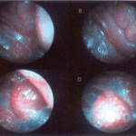
The following images were obtained by endoscopy of falcons with aspergillosis.A and B Lung Worm (S) over liver (Li) (serratospiculum seurati)C and D Aspergilloma (A) and prominent blood vessels on the caudal thoracic air sac (T).
