Date: 26 November 2013
Secondary metabolites, 3D structure: Trivial name – versicolorin A
Copyright: n/a
Notes:
Species: A. flavus, A. versicolorSystematic name; Anthra(2,3-b)furo(3,2-d)furan-5,10-dione, 3a,12a-dihydro-4,6,8-trihydroxy-, Z-(-)- Z-(-)-4,6,8-Trihydroxy-3a,12a-dihydroanthra(2,3-b)furo(3,2-d)furan-5,10-dione 4,6,8-Trihydroxy-3a,12a-dihydroanthra[2,3-b]furo[3,2-d]furan-5,10-dione Anthra[2,3-b]furo[3Molecular formulae: C18H10O7Molecular weight: 338.268Chemical abstracts number: 6807-96-1Selected references: Mori H, Kitamura J, Sugie S, Kawai K, Hamasaki T. Genotoxicity of fungal metabolites related to aflatoxin B1 biosynthesis. Mutat Res. 1985 Jul;143(3):121-5. Anderson MS, Dutton MF. Biosynthesis of versicolorin A. Appl Environ Microbiol. 1980 Oct;40(4):706-9.Toxicity: Little or no recorded toxicity in vertebrates but important as a representative of a group of metabolites which are precursors of the aflatoxins and sterigmatocystins.mouse LD50 intravenous 20mg/kg (20mg/kg) CRC Handbook of Antibiotic Compounds, Vols.1- , Berdy, J., Boca Raton, FL, CRC Press, 1980Vol. 3, Pg. 189, 1980. Bennett JW, Christensen SB. New perspectives on aflatoxin biosynthesis. Adv Appl Microbiol. 1983;29:53-92.
Images library
-
Title
Legend
-
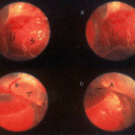
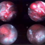
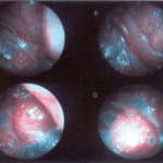
Falcons: The following images were obtained by endoscopy of falcons with aspergillosis.A,B Thoracic airsac (T) with prominent blood vessels and a dead serratospiculum worm (W). The presence of these lung worms makes the airsac look milky. D Normal ovary with developing follicles.

-
Falcons: The following images were obtained by endoscopy of falcons with aspergillosis.B,D Aspergillus lesions (A) over a swollen liver

-
Falcons: The following images were obtained by endoscopy of falcons with aspergillosis.B Cranial, middle, caudal lobes (K1,K2,K3) of the left kidney, all the lobes show slight nephromegaly.C Yellow aspergillus colony (A1), lying adjacent to the lung.D White aspergillus colonies (A2,A3,A4).
-
Falcons: The following images were obtained by endoscopy of falcons with aspergillosis.C Cranial pole of left kidney (K) -mildly inflamed.D Ovary ( F) with developing follicles.
-
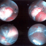
The following images were obtained by endoscopy of falcons with aspergillosis.A and B Lung Worm (S) over liver (Li) (serratospiculum seurati)C and D Aspergilloma (A) and prominent blood vessels on the caudal thoracic air sac (T).
