Date: 26 November 2013
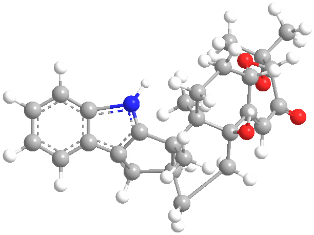
Secondary metabolites, 3D structure: Trivial name – Paxillin
Copyright: n/a
Notes:
Species: A. clavatoflavusSystematic name: 4b-Hydroxy-2-(1-hydroxy-1-methylethyl)-12b,12c-dimethyl-5,6,6a,7,12,12b,12c,13,14,14a-decahydro-2H-chromeno[5′,6′:6,7]indeno[1,2-b]indol-3(4bH)-oneMolecular formulae: C27H33NO4Molecular weight: 435.555Chemical abstracts number: 57186-25-1Selected references: http://www.pubmedcentral.nih.gov/ articlerender.fcgi?artid=525135
Images library
-
Title
Legend
-
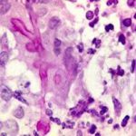
BAL specimen showing hyaline, septate hyphae consistent with Aspergillus, stained with Blankophor
-
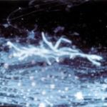
Mucous plug examined by light microscopy with KOH, showing a network of hyaline branching hyphae typical of Aspergillus, from a patient with ABPA.
-
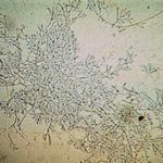
Corneal scraping stained with lactophenol cotton blue showing beaded septate hyphae not typical of either Fusarium spp or Aspergillus spp, being more consistent with a dematiceous (ie brown coloured) fungus
-
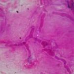
Corneal scrape with lactophenol cotton blue shows separate hyphae with Fusarium spp or Aspergillus spp.
-
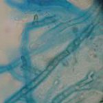
A filamentous fungus in the CSF of a patient with meningitis that grew Candida albicans in culture subsequently.
-
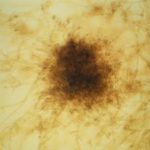
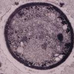
Transmission electron micrograph of a C. neoformans cell seen in CSF in an AIDS patients with remarkably little capsule present. These cells may be mistaken for lymphocytes.
-
India ink preparation of CSF showing multiple yeasts with large capsules, and narrow buds to smaller daughter cells, typical of C. neoformans
