Date: 26 November 2013
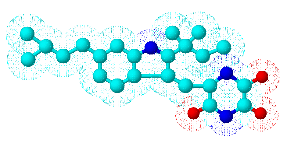
Secondary metabolites, 3D structure: Trivial name – neoechinuline
Copyright: n/a
Notes:
Species: A. amstelodamiSystematic name: (6E)-6-([2-(1,1-Dimethyl-2-propenyl)-6-(3-methyl-2-butenyl)-1H-indol-3-yl]methylene)-2,3,5-piperazinetrione 2,3,5-Piperazinetrione, 6-[[2-(1,1-dimethylallyl)-6-(3-methyl-2-butenyl)indol-3-yl]methylene]- Piperazinetrione, [[2-(1,1-dimethyl-2-propenyl)-6-Molecular formulae: C23H25N3O3Molecular weight: 391.463Chemical abstracts number: 25644-25-1Selected references: Selva A, Traldi P. Biomed Mass Spectrom. 1977 Jun;4(3):143-5. The electron impact induced fragmentation of Aspergillus amstelodami alkaloids and derivatives.
Images library
-
Title
Legend
-
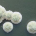
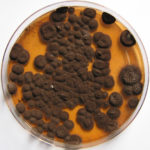
Pigmentation of Aspergillus versicolor colonies ranged from pale green to greenish-beige, pink-green, dark green and brown. Reverse is usually reddish. The growth rate is usually slow. Cultured on Sabouraud dextrose agar with chloramphenicol.
-
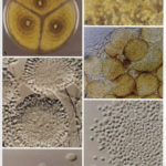
A Colonies on MEA after one week; B, C conidial heads with tip of conidiophire, x920; D conidial head, x 2330; E conidial heads x920
![aspvers[2] aspvers2](https://www.aspergillus.org.uk/wp-content/uploads/2017/10/aspvers2-150x150.jpg)
-
A Colonies on MEA + 20% sucrose after one week; B detail of colony showing columnar conidial heads x 44 ; C conidial heads x 920; D conidia x2330
-
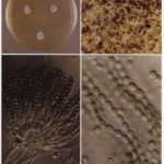
Cultures are grown on malt extract agar for 5-7 days at 30°C.
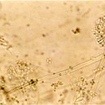
Light microscopy-1000x stained with lacto-phenol and cotton blue.
-
A Colonies on MEA +20% sucrose after one week; B ascomata x 40; C conidiophores x 920; D ascospores x2330; E ascoma x 230; F portion of ascoma with asci and ascospores, x 920.
