Date: 26 November 2013
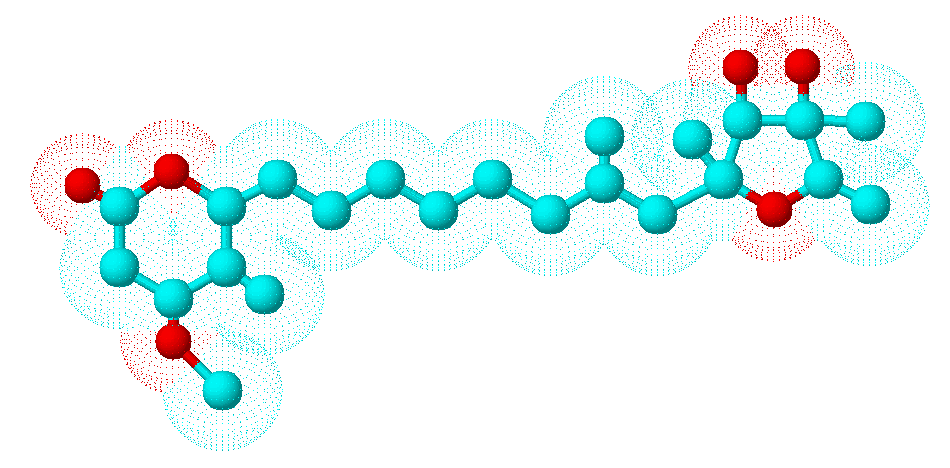
Secondary metabolites, 3D structure: Trivial name – citreoviridin
Copyright: n/a
Notes:
Species: A. terreusSystematic name: D-Iditol, 2,5-anhydro-1,6-dideoxy-2-C-[(1E,3E,5E,7E)-8-(4-methoxy-5-methyl-2-oxo-2H-pyran-6-yl)-2-methyl-1,3,5,7-octatetraenyl]-4-C-methyl- (9CI)Molecular formulae: C23H30O6Molecular weight: 402.481Chemical abstracts number: 25425-12-1Selected references: Franck B, Gehrken HP. Angew Chem Int Ed Engl. 1980;19(6):461-2 Citreoviridins from Aspergillus terreus.Toxicity: Citreoviridin is produced by P. citreonigrum (synonyms P. citreoviride and P. toxicarium), particularly in rice after harvest. It can cause cardiac beriberi in man. Acute cardiac beriberi in Japan is now only of historical interest although P. citreonigrum and citreoviridin are still reported in other parts of Asia. The fungus is said to be favoured by the lower temperatures and shorter hours of daylight occurring in the more temperate rice growing areas. The toxin is also produced by P. ochrosalmoneum. Citreoviridin has been found in un-harvested corn in the USA. Citreoviridin is an unusual molecule consisting of a lactone ring conjugated to a furan ring, with a molecular weight of 402. It is a neurotoxin. Nishie K, Cole RJ, Dorner JW. Res Commun Chem Pathol Pharmacol. 1988 Jan;59(1):31-52.Toxicity of citreoviridin.
Images library
-
Title
Legend
-
BAL specimen showing hyaline, septate hyphae consistent with Aspergillus, stained with Blankophor
-
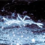
Mucous plug examined by light microscopy with KOH, showing a network of hyaline branching hyphae typical of Aspergillus, from a patient with ABPA.
-
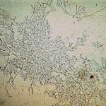
Corneal scraping stained with lactophenol cotton blue showing beaded septate hyphae not typical of either Fusarium spp or Aspergillus spp, being more consistent with a dematiceous (ie brown coloured) fungus
-
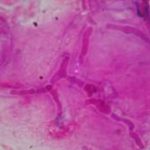
Corneal scrape with lactophenol cotton blue shows separate hyphae with Fusarium spp or Aspergillus spp.
-
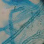
A filamentous fungus in the CSF of a patient with meningitis that grew Candida albicans in culture subsequently.
-
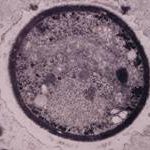
Transmission electron micrograph of a C. neoformans cell seen in CSF in an AIDS patients with remarkably little capsule present. These cells may be mistaken for lymphocytes.
-
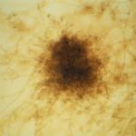
India ink preparation of CSF showing multiple yeasts with large capsules, and narrow buds to smaller daughter cells, typical of C. neoformans
