Date: 26 November 2013
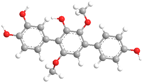
Secondary metabolites, 3D structure: Trivial name – 3-hydroxyterphenyllin
Copyright: n/a
Notes:
Species: A. candidusSystematic name: [1,1′:4′,1”-Terphenyl]-2′,3,4,4”-tetrol, 3′,6′-dimethoxy-Molecular formulae: C20H18O6Molecular weight: 354Chemical abstracts number: 66163-76-6PubChem URL: Go to PubChem linkSelected references: Kurobane, Itsuo; Vining, Leo C.; McInnes, A. Gavin; Smith, Donald G. (Dep. Biol., Dalhousie Univ., Halifax, NS B3H 4J1, Can.). J. Antibiot., 32(6), 559-64 (English) 1979.
Images library
-
Title
Legend
-
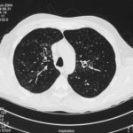
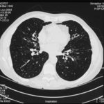
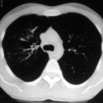
High resolution CT scan images with reconstruction of 1mm thick slices at approximately 10mm increments. The scan shows moderately severe multi-lobar cylindrical and varicose bronchiectasis predominantly centrally and in the upper lungs. There is no mucus plugging seen.
The features are in keeping with allergic bronchopulmonary aspergillosis
 ,
,  ,
, 
-
pt.SB – 6/10/98 – bronchocentric granulomatosis. CT scan showing multiple small nodules of variable size in both lung fields, apparently close to the vascular bundles.

-
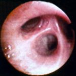
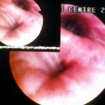
Bronchial oedema.Remarkably oedematous bronchial mucosa, as seen in ABPA.
-
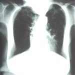
An example of longstanding allergic bronchopulmonary aspergillosis in a patient who has been steroid dependent for over 15 years showing remarkable kyphoscoliosis and honey combing and fibrosis of both lungs.
-
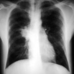
Recurrent pulmonary shadows 1. 6 Jan 1988 – chest radiograph showing right hilar enlargement, consistent with ABPA.
Recurrent pulmonary shadows 1. 3 Feb 1989 – chest radiograph showing right upper-lobe consolidation and contraction consistent with obstruction of RUL bronchus, in ABPA.
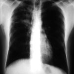
Clearing of pulmonary shadows 3, pt BJ. 5 April 1989 – resolution of shadows seen in February, with a course of corticosteroids.
Recurrence of pulmonary shadows 4, pt BJ. 2 September 1989 – recurrence of pulmonary shadows with an exacerbation of ABPA.
Central bronchiectasis, pt BJ. CT scan of thorax October 1989 showing central bronchiectasis, characteristic of ABPA (and cystic fibrosis).
 ,
,  ,
,  ,
,  ,
, 
-
A typical example of a wet mount of a sputum sample from a patient with allergic bronchopulmonary aspergillosis.


