Date: 26 November 2013
A Bronchogram showing saccular bronchiectasis
Copyright:
Fungal Infection Trust
Notes:
Bronchography (A & B)is an old technique for visualising the bronchial tree, by introducing radio-opaque dye into the airways and then taking a chest Xray. It was the first means used to diagnose bronchiectasis, seen in these examples below. An historical description of the technique from 1922 can be seen here
Nowadays CT scanning of the chest is used for this purpose with 3D reconstruction in some cases.
White cell scan (C) This pair of white cell scans from different people show almost no white cells in the lungs on the left, as in a healthy person (the spleen is the ‘hottest area). The scan on the right shows grossly increased update, especially in the left lung (seen on the right). This is the typical feature of severe bronchiectasis with large amounts of neutrophils and other phagocytes present.
Sinusitis Plain X-ray (D) of the face showing the maxillary sinuses. The right maxillary sinus is complete fluid filled and the left side (seen on the right) has a fluid level. These features may be seen with severe acute bacterial sinusitis, but also other causes of sinusitis, including allergic rhinosinusitis.
Images library
-
Title
Legend
-
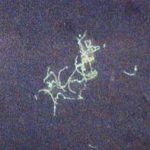
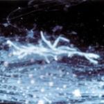
BAL specimen showing hyaline, septate hyphae consistent with Aspergillus, stained with Blankophor
-
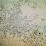
Mucous plug examined by light microscopy with KOH, showing a network of hyaline branching hyphae typical of Aspergillus, from a patient with ABPA.
-
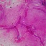
Corneal scraping stained with lactophenol cotton blue showing beaded septate hyphae not typical of either Fusarium spp or Aspergillus spp, being more consistent with a dematiceous (ie brown coloured) fungus
-
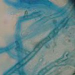
Corneal scrape with lactophenol cotton blue shows separate hyphae with Fusarium spp or Aspergillus spp.
-
A filamentous fungus in the CSF of a patient with meningitis that grew Candida albicans in culture subsequently.
-
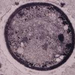
Transmission electron micrograph of a C. neoformans cell seen in CSF in an AIDS patients with remarkably little capsule present. These cells may be mistaken for lymphocytes.
-
India ink preparation of CSF showing multiple yeasts with large capsules, and narrow buds to smaller daughter cells, typical of C. neoformans
