Date: 6 November 2014
Copyright: n/a
Notes:
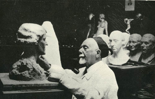
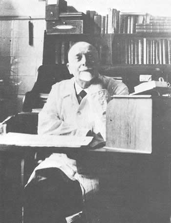
In addition to being a worldwide known mycologist, Sabouraud was a talented painter and sculptor. Short biography and bibliography of Sabouraud
The outstanding names in the history of any branch of science are not always those of the men who made the primary observation. The prevailing climate of opinion and the available techniques greatly affect the contemporary significance attributed to any novel discovery and the historical land marks are frequently the reputations of subsequent workers who were men of their time, who showed singleness of purpose and who crystallise ideas which were nearing supersaturation. Raimond Sabauroud the French Dermatologist, was such a man. In the early 1890’s by remaking observations which had been on record for fifty years but not universally accepted, he was able to silence finally the view that the association of fungi with ringworm was incidental.
Using the recently developed pure culture techniques he was able to establish convincingly the plurality of the ringworm fungi, and his medical training enabled him to integrate the mycological and clinical aspects of ringworm. He was stimulated by the advances made by able contemporaries in both medicine and veterinary science and finally in 1910 he codified both his own and their results in the monumental Les Teignes, one of the most comprehensive treatments ever given to a group of pathogenic fungi and a monograph to which students of the dermataophytes must still refer.
The dermatomycoses have always been a basic theme of medical and veterinary mycology and at the turn of the century they overshadowed the systemic mycoses by which the dermatomycoses are overshadowed today. They served to focus attention on fungi as pathogens of man and higher animals and provided a seed from which an interesting branch of medicine and vet science has emerged.
From Sabouraudia vol 1 1961-2 p1 by GC Ainsworth
Images library
-
Title
Legend
-
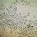
Mucous plug examined by light microscopy with KOH, showing a network of hyaline branching hyphae typical of Aspergillus, from a patient with ABPA.
-
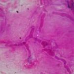
Corneal scraping stained with lactophenol cotton blue showing beaded septate hyphae not typical of either Fusarium spp or Aspergillus spp, being more consistent with a dematiceous (ie brown coloured) fungus
-
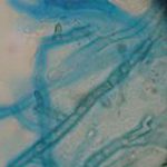
Corneal scrape with lactophenol cotton blue shows separate hyphae with Fusarium spp or Aspergillus spp.
-
A filamentous fungus in the CSF of a patient with meningitis that grew Candida albicans in culture subsequently.
-
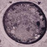
Transmission electron micrograph of a C. neoformans cell seen in CSF in an AIDS patients with remarkably little capsule present. These cells may be mistaken for lymphocytes.
-
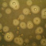
India ink preparation of CSF showing multiple yeasts with large capsules, and narrow buds to smaller daughter cells, typical of C. neoformans
-
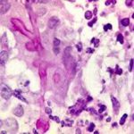
PAS stain. An example of Aspergillus fumigatus.
(PAS-stained) in a patient with chronic granulomatous disease showing a 45 degree branching hypha within a giant cell. Rather bulbous hyphal ends are also seem, which is sometimes found inAspergillus spp. infections, histologically. (x800)

