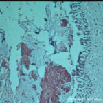
Date: 26 November 2013
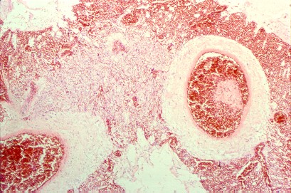
Pulmonary aspergillosis (K&E) (parrot C). Tissue from an individually housed and recently purchased, 6 month old African grey parrot found dead in the cage. Necropsy examination revealed focal necrosis of the left lung. This section stained by haematoxylin and eosin reveals septate fungal hyphae within the lung parenchyma. Similar hyphae were located in the walls and lumen of parabronchi, and within the walls of pulmonary blood vessels.
Copyright:
© Dr. Michael Day, University of Bristol
Notes: n/a
Images library
-
Title
Legend
-
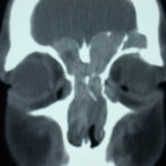
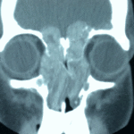
4 Total obstruction of the sinuses due to inflamed mucosa. (Patient 04)
-
1 Axial computed tomography (CT) scans of the frontal sinus.
A: due to the long lasting pressure of mucus, the bone of the anterior wall of frontal sinus is thinned out and elevated anteriorly, forming a bulge. B: same situation as depicted in fig A: the posterior bony wall of frontal sinus is thinned out and extremely elevated posteriorly towards the frontal lobe of the brain. As depicted on the scan, a thin bony layer covering the dura could be recognized intraoperatively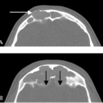
-
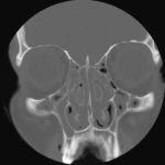
2 Same patient as 1 and 3, frontal CT
-
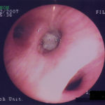
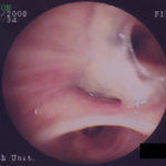
D. 6 months later, tenacious yellow secretions in L basal bronchial division
-
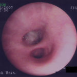
C. After suction the material was seen to extend distally – obstructing the right basal stem bronchus
-
B. After suction the material was seen to extend distally – obstructing the right basal stem bronchus
